GFAP Antibody (5C10)
Novus Biologicals, part of Bio-Techne | Catalog # NBP1-05197


Conjugate
Catalog #
Key Product Details
Species Reactivity
Validated:
Human, Mouse, Rat, Porcine, Bovine, Equine
Cited:
Human, Mouse, Rat, Porcine
Applications
Validated:
Immunocytochemistry/Immunofluorescence, Immunohistochemistry, Immunohistochemistry Free-Floating, Immunohistochemistry-Frozen, Immunohistochemistry-Paraffin, Simple Western, Western Blot
Cited:
Block/Neutralize, IF/IHC, IHC/IF, Immunocytochemistry/ Immunofluorescence, Immunohistochemistry, Immunohistochemistry-Frozen, Immunohistochemistry-Paraffin, Western Blot
Label
Unconjugated
Antibody Source
Monoclonal Mouse IgG1 Clone # 5C10
Concentration
1.0 mg/ml
Product Summary for GFAP Antibody (5C10)
Immunogen
This GFAP Antibody (5C10) was developed against a preparation of purified pig spinal cord GFAP
Localization
Cytoplasm. Note: Associated with intermediate filaments.
Marker
Astrocyte Marker
Clonality
Monoclonal
Host
Mouse
Isotype
IgG1
Theoretical MW
50 kDa.
Disclaimer note: The observed molecular weight of the protein may vary from the listed predicted molecular weight due to post translational modifications, post translation cleavages, relative charges, and other experimental factors.
Disclaimer note: The observed molecular weight of the protein may vary from the listed predicted molecular weight due to post translational modifications, post translation cleavages, relative charges, and other experimental factors.
Scientific Data Images for GFAP Antibody (5C10)
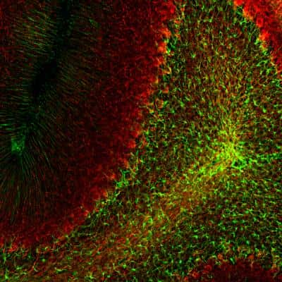
Immunohistochemistry Free-Floating: GFAP Antibody (5C10) [NBP1-05197] - Analysis of rat cerebellum section stained with mouse GFAP mAb, dilution 1:1,000 (Green), costained with rabbit neurofilament NF-L pAb, dilution 1:2,000 (Red). Following transcardial perfusion with 4% paraformaldehyde, brain was post fixed for 24hrs, cut to 45uM, and free-floating sections were stained with antibodies. The GFAP antibody stains a network of astroglial cells, while the NF-L antibody labels neuronal cells and their processes.

Simple Western: GFAP Antibody (5C10) [NBP1-05197] - Simple Western lane view shows a specific band for GFAP in 0.05 mg/ml of Human Brain lysate. This experiment was performed under reducing conditions using the 12-230 kDa separation system.

Immunohistochemistry: GFAP Antibody (5C10) [NBP1-05197] - The immunofluorescence of TLR7 recognized by Alexa 488, green (NBP2-24906). GFAP recognized by Alexa 594, red. NEUN recognized by Alexa 633, (blue) and merged image in the hippocampal region. NeuN and GFAP were applied to show the distribution of TLR7 within neuronal and supportive tissue populations. Scale bar 80 um. Image collected and cropped by CiteAb from the following publication (//doi.org/10.1371/journal.pone.0222818) licensed under a CC-BY license.
Applications for GFAP Antibody (5C10)
Application
Recommended Usage
Immunocytochemistry/Immunofluorescence
1:1000
Immunohistochemistry
1:1000
Immunohistochemistry Free-Floating
1:1000
Immunohistochemistry-Frozen
1:1000
Immunohistochemistry-Paraffin
1:1000
Simple Western
1:3000
Western Blot
1:5000
Application Notes
This GFAP (5C10) antibody is useful for Immunocytochemistry/Immunofluorescence, Immunohistochemistry on paraffin-embedded and frozen sections, and Western blot. In WB, a band can be seen at approx. 50 kDa.
In Simple Western only 10 - 15 uL of the recommended dilution is used per data point. Separated by Size-Wes, Sally Sue/Peggy Sue.
In Simple Western only 10 - 15 uL of the recommended dilution is used per data point. Separated by Size-Wes, Sally Sue/Peggy Sue.
Please Note: Optimal dilutions of this antibody should be experimentally determined.
Reviewed Applications
Read 1 review rated 3 using NBP1-05197 in the following applications:
Formulation, Preparation, and Storage
Purification
Immunogen affinity purified
Formulation
PBS, 50% glycerol
Preservative
5mM Sodium Azide
Concentration
1.0 mg/ml
Shipping
The product is shipped with polar packs. Upon receipt, store it immediately at the temperature recommended below.
Stability & Storage
Store at 4C short term. Aliquot and store at -20C long term. Avoid freeze-thaw cycles.
Background: GFAP
An increase in GFAP levels is often associated with neuroinflammation which results in the activation and proliferation of astroglia cell population (1,2). GFAP expression is also observed in brains of patients with neurodegenerative diseases including Alzheimer's and Parkinson's, epilepsy disorders, and brain injuries (1-4). Lesion sites associated with neurodegeneration can exhibit an array of gliosis characteristics from glial scarring with reduced astrocyte proliferation to activated, GFAP-positive astrocytes surrounding amyloid plaques (2). Furthermore, the GFAP gene is a target of single nucleotide polymorphisms in the coding region, considered a gain-of-function mutation, characterized by astrocytic inclusions, termed Rosenthal fibers, resulting in Alexander Disease (1-4). GFAP is also a center of many post-translational modifications, such as phosphorylation, which can alter various aspects of filament assembly (1,4).
References
1. Yang, Z., & Wang, K. K. (2015). Glial fibrillary acidic protein: from intermediate filament assembly and gliosis to neurobiomarker. Trends in Neurosciences. https://doi.org/10.1016/j.tins.2015.04.003
2. Hol, E. M., & Capetanaki, Y. (2017). Type III Intermediate Filaments Desmin, Glial Fibrillary Acidic Protein (GFAP), Vimentin, and Peripherin. Cold Spring Harbor Perspectives in Biology. https://doi.org/10.1101/cshperspect.a021642
3. Potokar, M., Morita, M., Wiche, G., & Jorgacevski, J. (2020). The Diversity of Intermediate Filaments in Astrocytes. Cells. https://doi.org/10.3390/cells9071604
4. Viedma-Poyatos, a., Pajares, M. A., & Perez-Sala, D. (2020). Type III intermediate filaments as targets and effectors of electrophiles and oxidants. Redox Biology. https://doi.org/10.1016/j.redox.2020.101582
Long Name
Glial Fibrillary Acidic Protein
Alternate Names
ALXDRD, FLJ45472, GFAP, GFAP astrocytes, glial fibrillary acidic protein
Gene Symbol
GFAP
Additional GFAP Products
Product Documents for GFAP Antibody (5C10)
Product Specific Notices for GFAP Antibody (5C10)
This product is for research use only and is not approved for use in humans or in clinical diagnosis. Primary Antibodies are guaranteed for 1 year from date of receipt.
Loading...
Loading...
Loading...
Loading...