Human alpha-Galactosidase A/GLA Antibody Best Seller
R&D Systems, part of Bio-Techne | Catalog # AF6146

Key Product Details
Species Reactivity
Validated:
Cited:
Applications
Validated:
Cited:
Label
Antibody Source
Product Specifications
Immunogen
Leu32-Leu429
Accession # P06280
Specificity
Clonality
Host
Isotype
Scientific Data Images for Human alpha-Galactosidase A/GLA Antibody
Detection of Human alpha‑Galactosidase A/GLA by Western Blot.
Western blot shows lysates of A549 human lung carcinoma cell line, HeLa human cervical epithelial carcinoma cell line, and HepG2 human hepatocellular carcinoma cell line. PVDF membrane was probed with 2 µg/mL of Sheep Anti-Human a-Galactosidase A/GLA Antigen Affinity-purified Polyclonal Antibody (Catalog # AF6146) followed by HRP-conjugated Anti-Sheep IgG Secondary Antibody (Catalog # HAF016). A specific band was detected for a-Galactosidase A/GLA at approximately 38 kDa (as indicated). This experiment was conducted under reducing conditions and using Immunoblot Buffer Group 1.alpha‑Galactosidase A/GLA in HeLa Human Cell Line.
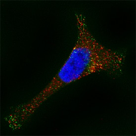
a-Galactosidase A/GLA was detected in immersion fixed HeLa human cervical epithelial carcinoma cell line using Sheep Anti-Human a-Galactosidase A/GLA Antigen Affinity-purified Polyclonal Antibody (Catalog # AF6146) at 15 µg/mL for 3 hours at room temperature. Cells were stained using the Northern-Lights™ 557-conjugated Anti-Sheep IgG Secondary Antibody (red; Catalog # NL010). LAMP1 was also detected using Mouse Anti-Human LAMP1 Mono-clonal Antibody (Catalog # MAB4800). Cells were stained using the Northern-Lights™ 493-conjugated Anti-Mouse IgG Secondary Antibody (green; Catalog # NL009). Cells were counterstained with DAPI (blue). Specific staining was localized to lysosomes. View our protocol for Fluorescent ICC Staining of Cells on Coverslips.Detection of Human alpha‑Galactosidase A/GLA by Simple WesternTM.
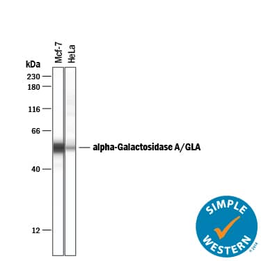
Simple Western lane view shows lysates of MCF-7 human breast cancer cell line and HeLa human cervical epithelial carcinoma cell line, loaded at 0.2 mg/mL. A specific band was detected for a-Galactosidase A/GLA at approximately 55 kDa (as indicated) using 50 µg/mL of Sheep Anti-Human a-Galactosidase A/GLA Antigen Affinity-purified Polyclonal Antibody (Catalog # AF6146) followed by 1:50 dilution of HRP-conjugated Anti-Sheep IgG Secondary Antibody (Catalog # HAF016). This experiment was conducted under reducing conditions and using the 12-230 kDa separation system.Applications for Human alpha-Galactosidase A/GLA Antibody
Immunocytochemistry
Sample: Immersion fixed HeLa human cervical epithelial carcinoma cell line
Simple Western
Sample: MCF‑7 human breast cancer cell line and HeLa human cervical epithelial carcinoma cell line
Western Blot
Sample: A549 human lung carcinoma cell line, HeLa human cervical epithelial carcinoma cell line, and HepG2 human hepatocellular carcinoma cell line
Formulation, Preparation, and Storage
Purification
Reconstitution
Formulation
Shipping
Stability & Storage
- 12 months from date of receipt, -20 to -70 °C as supplied.
- 1 month, 2 to 8 °C under sterile conditions after reconstitution.
- 6 months, -20 to -70 °C under sterile conditions after reconstitution.
Background: alpha-Galactosidase A/GLA
Human alpha-Galactosidase A is a homodimeric glycoprotein that can release terminal alpha-galactosyl moieties from glycolipids and glycoproteins and catalyze the hydrolysis of melibiose into galactose and glucose (1). It is a lysosomal enzyme and is responsible for degradation of glycolipid globotriaosylceramide (Gb3) (Gal alpha1‑4Gal beta1‑4Glc beta‑ceramide). Mutations in this gene cause Fabry disease, an X-linked hereditary lysosomal storage disease with the accumulation of Gb3 in the walls of small blood vessels, nerves, dorsal root ganglia, renal glomerular and tubular epithelial cells, and cardiomyocytes (2, 3). Inability to prevent the glycosphingolipid deposition can cause hypertension, strokes, heart attack and progressive renal failure (4). Current treatment for Fabry disease is enzyme replacement therapy using intravenously delivered recombinant alpha-Galactosidase A (5, 6).
References
-
Ioannou, Y.A. et al. (1998) Biochem. J. 332:789.
-
Koide, T. et al. (1990) FEBS Lett. 259:353.
-
Ioannou Y.A, et al. (1992) J. Cell Biol. 119:1137.
-
Germain, D.P. (2002) Expert. Opin. Investig. Drugs. 11:1467.
-
Barngrover, D. (2003) J. Biotechnol. 95:280.
-
Mignani, R. and Cagnoli, L. (2004) J. Nephrol. 17:354.
Alternate Names
Entrez Gene IDs
Gene Symbol
UniProt
Additional alpha-Galactosidase A/GLA Products
Product Documents for Human alpha-Galactosidase A/GLA Antibody
Product Specific Notices for Human alpha-Galactosidase A/GLA Antibody
For research use only


