Human NCAM-1/CD56 PE-conjugated Antibody
R&D Systems, part of Bio-Techne | Catalog # FAB2408P


Key Product Details
Species Reactivity
Validated:
Cited:
Applications
Validated:
Cited:
Label
Antibody Source
Product Specifications
Immunogen
Specificity
Clonality
Host
Isotype
Scientific Data Images for Human NCAM-1/CD56 PE-conjugated Antibody
Detection of NCAM‑1/CD56 in Human PBMCs by Flow Cytometry.
Human peripheral blood mononuclear cells (PBMCs) were stained with Mouse Anti-Human CD3e Alexa Fluor® 405-conjugated Monoclonal Antibody (Catalog # FAB100V) and either (A) Mouse Anti-Human NCAM-1/CD56 PE-conjugated Monoclonal Antibody (Catalog # FAB2408P) or (B) Mouse IgG2BPhycoerythrin Isotype Control (Catalog # IC0041P). View our protocol for Staining Membrane-associated Proteins.Detection of Human NCAM-1/CD56 by Flow Cytometry
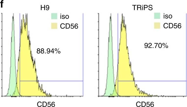
Characterization of iMPCs during monolayer differentiation. a–e Representative immunostaining of Pax3 (a), Myf5 (b), MyoD (c), and MyoG (d), and corresponding quantification (e) during iMPC expansion. Scale bar=100 µm. f Representative FACS analysis for CD56 in H9 and TRiPSC derived iMPCs. g Representative immunostaining (top) and quantification (bottom) of Pax7+ and MyoG+ cell populations for H9 and TRiPS derived myotubes at 2 weeks of monolayer differentiation. (n = 6 samples from 2 differentiations for each cell line). h Representative immunostaining and quantification of GFP+/Pax7+ and GFP-/Pax7+ cell pools at 2 weeks of monolayer differentiation. Scale bar=50 µm. (n = 4 samples from 2 differentiations for each cell line). i Representative immunostaining and quantification of myotube diameter at 1, 2, and 4 weeks of monolayer differentiation. (*P < 0.05 vs. 1 week, #P < 0.05 vs. 4 week, Tukey–Kramer HSD test; n = 6 samples from 2 differentiations for each cell line). Scale bars=50 µm. Data are presented as mean ± SEM Image collected and cropped by CiteAb from the following publication (https://pubmed.ncbi.nlm.nih.gov/29317646), licensed under a CC-BY license. Not internally tested by R&D Systems.Applications for Human NCAM-1/CD56 PE-conjugated Antibody
Flow Cytometry
Sample: Human peripheral blood mononuclear cells (PBMCs)
Reviewed Applications
Read 1 review rated 4 using FAB2408P in the following applications:
Formulation, Preparation, and Storage
Purification
Formulation
Shipping
Stability & Storage
- 12 months from date of receipt, 2 to 8 °C as supplied.
Background: NCAM-1/CD56
Neural cell adhesion molecule 1 (NCAM-1) is a multifunctional member of the Ig superfamily. It belongs to a family of membrane-bound glycoproteins that are involved in Ca++ independent cell matrix and homophilic or heterophilic cell-cell interactions. NCAM-1 specifically binds to heparan sulfate proteoglycans (1), the extracellular matrix protein agrin (2), and several chondroitin sulfate proteoglycans that include neurocan and phosphocan (3). There are three main forms of human NCAM-1 that arise by alternate splicing. These are designated NCAM-120/NCAM-1 (761 amino acids [aa]), NCAM-140 (848 aa), and NCAM-180 (1120 aa). NCAM-120 is GPI-linked, while NCAM-140 and NCAM-180 are type I transmembrane glycoproteins (4‑6). Additional alternate splicing adds considerable diversity to all three forms, and extracellular proteolytic processing is possible for NCAM-180 (7‑8). NCAM-1 is synthesized as a 761 aa preproprecursor that contains a 19 aa signal sequence, a 722 aa GPI-linked mature region, and a 20 aa C-terminal prosegment (4). The molecule contains five C-2 type Ig-like domains and two fibronectin type-III domains. Human to mouse, NCAM-1 is 93% aa identical. NCAM-1 appears to be highly sialylated. The polysialyation of NCAM-1 reduces its adhesive property and increases its neurite outgrowth promoting features (9). NCAM-1 in the adult brain shows a decline of sialylation relative to earlier developmental periods. In regions that retain a high degree of neuronal plasticity, however, the adult brain continues to express polysialylation-NCAM-1, suggesting sialylation of NCAM-1 is involved in regenerative processes and synaptic plasticity (10‑13).
References
- Burg, M.A. et al. (1995) J. Neurosci. Res. 41:49.
- Storms, S.D. and U. Rutishauser (1998) J Biol. Chem. 273:27124.
- Margolis, R.K. et al. (1996) Perspect. Dev. Neurobiol. 3:273.
- Dickson, G. et al. (1987) Cell 50:1119.
- Lanier, L.L. et al. (1991) J. Immunol. 146:4421.
- Hemperly, J.J. et al. (1990) J. Mol. Neurosci. 2:71.
- Rutishauser, U.and C. Goridis (1986) Trends Genet. 2:72.
- Vawter, M.P. et al. (2001) Exp. Neurol. 172:29.
- Rutihauser, U. (1990) Adv. Exp. Med. Biol. 265:179.
- Becker, C.G. et al. (1996) J. Neurosci. Res. 45:143.
- Doherty, P. et al. (1995) J. Neurobiol. 26:437.
- Eckardt, M. et al. (2000) J. Neurosci. 20:5234.
- Muller, D. et al. (1996) Neuron 17:413.
Long Name
Alternate Names
Gene Symbol
Additional NCAM-1/CD56 Products
Product Documents for Human NCAM-1/CD56 PE-conjugated Antibody
Product Specific Notices for Human NCAM-1/CD56 PE-conjugated Antibody
For research use only
