Mouse Aminopeptidase N/CD13 Antibody
R&D Systems, part of Bio-Techne | Catalog # AF2335


Key Product Details
Species Reactivity
Validated:
Cited:
Applications
Validated:
Cited:
Label
Antibody Source
Product Summary for Mouse Aminopeptidase N/CD13 Antibody
Immunogen
Lys69-Ser966
Accession # P97449
Specificity
Clonality
Host
Isotype
Scientific Data Images for Mouse Aminopeptidase N/CD13 Antibody
Aminopeptidase N/CD13 in Mouse Splenocytes.
Aminopeptidase N/CD13 was detected in immersion fixed mouse splenocytes using Goat Anti-Mouse Aminopeptidase N/CD13 Antigen Affinity-purified Polyclonal Antibody (Catalog # AF2335) at 15 µg/mL for 3 hours at room temperature. Cells were stained using the NorthernLights™ 557-conjugated Anti-Goat IgG Secondary Antibody (red; Catalog # NL001) and counterstained with DAPI (blue). Specific staining was localized to cytoplasm. View our protocol for Fluorescent ICC Staining of Non-adherent Cells.Aminopeptidase N/CD13 in Mouse Kidney.
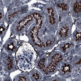
Aminopeptidase N/CD13 was detected in immersion fixed paraffin-embedded sections of mouse kidney using Goat Anti-Mouse Aminopeptidase N/CD13 Antigen Affinity-purified Polyclonal Antibody (Catalog # AF2335) at 0.1 µg/mL for 1 hour at room temperature followed by incubation with the Anti-Goat IgG VisUCyte™ HRP Polymer Antibody (Catalog # VC004). Before incubation with the primary antibody, tissue was subjected to heat-induced epitope retrieval using Antigen Retrieval Reagent-Basic (Catalog # CTS013). Tissue was stained using DAB (brown) and counterstained with hematoxylin (blue). Specific staining was localized to apical membrane. View our protocol for IHC Staining with VisUCyte HRP Polymer Detection Reagents.Applications for Mouse Aminopeptidase N/CD13 Antibody
CyTOF-ready
Flow Cytometry
Sample: bEnd.3 mouse endothelioma cell line
Immunocytochemistry
Sample: Immersion fixed mouse splenocytes
Immunohistochemistry
Sample: Immersion fixed paraffin-embedded sections of mouse kidney
Immunoprecipitation
Sample: Conditioned cell culture medium spiked with Recombinant Mouse Aminopeptidase N/CD13 (Catalog # 2335-ZN), see our available Western blot detection antibodies
Western Blot
Sample: Recombinant Mouse Aminopeptidase N/CD13 (Catalog # 2335-ZN)
Reviewed Applications
Read 1 review rated 5 using AF2335 in the following applications:
Formulation, Preparation, and Storage
Purification
Reconstitution
Formulation
Shipping
Stability & Storage
- 12 months from date of receipt, -20 to -70 °C as supplied.
- 1 month, 2 to 8 °C under sterile conditions after reconstitution.
- 6 months, -20 to -70 °C under sterile conditions after reconstitution.
Background: Aminopeptidase N/CD13
The mouse Anpep gene encodes Aminopeptidase N (APN), which is also known as microsomal aminopeptiase, alanyl aminopeptidase, Aminopeptidase M, CD13, or membrane protein p161 (1‑3). The deduced amino acid sequence of mouse APN consists of a short cytoplasmic tail (residues 2 to 8), a transmembrane region (residue 9 to 32), a Ser/Thr rich region and a zinc metalloprotease domain (residues 69 to 966). Widely expressed in many cells, tissues and species, APN cleaves the N-terminal amino acids from bioactive peptides, leading to their inactivation or degradation. The roles of APN in many fields, such as neuroscience, hematopoeitic cells, immune system, angiogenesis, cancer and viral infection, have been reviewed (4).
References
- Chen, H. et al. (1996) J. Immunol. 157:2593.
- Larsen, S.L. et al. (1996) J. Exp. Med. 184:183.
- Hansen, A.S. et al. (1993) Eur. J. Immunol. 23:2358.
- Turner, A.J. (2004) in Handbook of Proteolytic Enzymes (ed. Barrett, et al.) p. 289 Academic Press, San Diego.
Alternate Names
Gene Symbol
UniProt
Additional Aminopeptidase N/CD13 Products
Product Documents for Mouse Aminopeptidase N/CD13 Antibody
Product Specific Notices for Mouse Aminopeptidase N/CD13 Antibody
For research use only
