CD63 Antibody (H5C6) - BSA Free
Novus Biologicals, part of Bio-Techne | Catalog # NBP2-42225
Clone H5C6 was used by HLDA to establish CD designation.


Conjugate
Catalog #
Forumulation
Catalog #
Key Product Details
Species Reactivity
Validated:
Human, Canine
Cited:
Human, Mouse, Rat, Canine
Applications
Validated:
Block/Neutralize, Dot Blot, Electron Microscopy, ELISA, Flow (Intracellular), Flow Cytometry, Functional, Immunocytochemistry/Immunofluorescence, Immunohistochemistry Whole-Mount, Immunoprecipitation, In vitro assay, Western Blot
Cited:
Block/Neutralize, Cytometric Bead Assay Standard, Dot Blot, Electron Microscopy, ELISA, Flow Cytometry, Functional Assay, Immunoassay, Immunocytochemistry/ Immunofluorescence, Immunoprecipitation, In vitro assay, Western Blot
Label
Unconjugated
Antibody Source
Monoclonal Mouse IgG1 kappa Clone # H5C6
Format
BSA Free
Concentration
1.0 mg/ml
Product Summary for CD63 Antibody (H5C6) - BSA Free
Immunogen
Human splenic adherent cells.
Marker
Exosome Marker
Clonality
Monoclonal
Host
Mouse
Isotype
IgG1 kappa
Scientific Data Images for CD63 Antibody (H5C6) - BSA Free
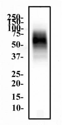
Western Blot: CD63 Antibody (H5C6) [NBP2-42225] - THP1 whole cell protein was separated by SDS-PAGE on a 12% gel and transferred to PVDF membrane. The membrane was probed with anti-CD63 antibody at 2 ug/mL and detected with an anti-mouse HRP secondary antibody using chemiluminescence.

Immunocytochemistry/Immunofluorescence: CD63 Antibody (H5C6) [NBP2-42225] - The CD63 (H5C6) antibody was tested in HeLa cells at a 1:50 dilution against DyLight 488 (Green). Actin and nuclei were counterstained against Phalloidin 568 (Red) and DAPI (Blue), respectively.

Immunocytochemistry/Immunofluorescence: CD63 Antibody (H5C6) [NBP2-42225] - HeLa cells were fixed in 4% paraformaldehyde for 10 minutes and permeabilized in 0.05% Triton X-100 in PBS for 5 minutes. The cells were incubated with anti-CD63 Antibody [H5C6] conjugated to DyLight 550 (NBP2-42225R) at 5 ug/ml for 1 hour at room temperature. Nuclei were counterstained with DAPI (Blue). Cells were imaged using a 100X objective and digitally deconvolved.


Applications for CD63 Antibody (H5C6) - BSA Free
Application
Recommended Usage
Block/Neutralize
reported in scientific literature (PMID 35602933)
Electron Microscopy
reported in scientific literature (PMID 16735575)
Flow Cytometry
1:1000
Functional
reported in scientific literature (PMID 9811687)
Immunocytochemistry/Immunofluorescence
1:50-1:100
Immunohistochemistry Whole-Mount
reported in scientific literature (PMID 21464080)
In vitro assay
reported in scientific literature (PMID 21464080)
Please Note: Optimal dilutions of this antibody should be experimentally determined.
Reviewed Applications
Read 5 reviews rated 4 using NBP2-42225 in the following applications:
Formulation, Preparation, and Storage
Purification
Protein G purified
Formulation
PBS
Format
BSA Free
Preservative
0.05% Sodium Azide
Concentration
1.0 mg/ml
Shipping
The product is shipped with polar packs. Upon receipt, store it immediately at the temperature recommended below.
Stability & Storage
Store at 4C short term. Aliquot and store at -20C long term. Avoid freeze-thaw cycles.
Background: CD63
References
1. Pols, M. S., & Klumperman, J. (2009). Trafficking and function of the tetraspanin CD63. Experimental cell research. https://doi.org/10.1016/j.yexcr.2008.09.020
2. Metzelaar, M. J., Wijngaard, P. L., Peters, P. J., Sixma, J. J., Nieuwenhuis, H. K., & Clevers, H. C. (1991). CD63 antigen. A novel lysosomal membrane glycoprotein, cloned by a screening procedure for intracellular antigens in eukaryotic cells. The Journal of biological chemistry.
3. Horejsi, V., & Vlcek, C. (1991). Novel structurally distinct family of leucocyte surface glycoproteins including CD9, CD37, CD53 and CD63. FEBS letters. https://doi.org/10.1016/0014-5793(91)80988-f
4. Eckfeld, C., HauBler, D., Schoeps, B., Hermann, C. D., & Kruger, A. (2019). Functional disparities within the TIMP family in cancer: hints from molecular divergence. Cancer metastasis reviews. https://doi.org/10.1007/s10555-019-09812-6
5. Hoffmann, H. J., Santos, A. F., Mayorga, C., Nopp, A., Eberlein, B., Ferrer, M., Rouzaire, P., Ebo, D. G., Sabato, V., Sanz, M. L., Pecaric-Petkovic, T., Patil, S. U., Hausmann, O. V., Shreffler, W. G., Korosec, P., & Knol, E. F. (2015). The clinical utility of basophil activation testing in diagnosis and monitoring of allergic disease. Allergy. https://doi.org/10.1111/all.12698
6. Dell'Angelica, E. C., Shotelersuk, V., Aguilar, R. C., Gahl, W. A., & Bonifacino, J. S. (1999). Altered trafficking of lysosomal proteins in Hermansky-Pudlak syndrome due to mutations in the beta 3A subunit of the AP-3 adaptor. Molecular cell. https://doi.org/10.1016/s1097-2765(00)80170-7
Alternate Names
CD63, Granulophysin, Lamp-3, ME491, OMA81H, Tspan30
Entrez Gene IDs
967 (Human)
Gene Symbol
CD63
UniProt
Additional CD63 Products
Product Documents for CD63 Antibody (H5C6) - BSA Free
Product Specific Notices for CD63 Antibody (H5C6) - BSA Free
This product is for research use only and is not approved for use in humans or in clinical diagnosis. Primary Antibodies are guaranteed for 1 year from date of receipt.
Loading...
Loading...
Loading...
Loading...
Loading...