HIF-1 alpha Antibody (H1alpha67) [Alexa Fluor® 647]
Novus Biologicals, part of Bio-Techne | Catalog # NB100-105AF647

Conjugate
Catalog #
Forumulation
Catalog #
Key Product Details
Species Reactivity
Validated:
Human, Mouse, Rat, Porcine, Bovine, Canine, Feline, Ferret, Monkey, Primate, Rabbit, Sheep, Xenopus
Cited:
Mouse
Applications
Validated:
ELISA, Flow Cytometry, Immunoblotting, Immunocytochemistry/Immunofluorescence, Immunohistochemistry, Immunohistochemistry-Frozen, Immunohistochemistry-Paraffin, Knockdown Validated, Knockout Validated, Product Image, Western Blot
Cited:
Flow Cytometry
Label
Alexa Fluor 647 (Excitation = 650 nm, Emission = 668 nm)
Antibody Source
Monoclonal Mouse IgG2B Clone # H1alpha67
Concentration
Please see the vial label for concentration. If unlisted please contact technical services.
Product Summary for HIF-1 alpha Antibody (H1alpha67) [Alexa Fluor® 647]
Immunogen
This HIF-1 alpha Antibody (H1alpha67) was developed against a fusion protein containing amino acids 432 - 528 of human HIF-1 alpha [Uniprot# Q16665].
Reactivity Notes
Candida Albicans reactivity was reported by customer review. Xenopus reactivity was reported in scientific literature (PMID: 18303027). Please note that this antibody is reactive to Mouse and derived from the same host, Mouse. Additional Mouse on Mouse blocking steps may be required for IHC and ICC experiments. Please contact Technical Support for more information.
Localization
HIF-1 is a nuclear protein
Clonality
Monoclonal
Host
Mouse
Isotype
IgG2B
Theoretical MW
93 kDa.
Disclaimer note: The observed molecular weight of the protein may vary from the listed predicted molecular weight due to post translational modifications, post translation cleavages, relative charges, and other experimental factors.
Disclaimer note: The observed molecular weight of the protein may vary from the listed predicted molecular weight due to post translational modifications, post translation cleavages, relative charges, and other experimental factors.
Scientific Data Images for HIF-1 alpha Antibody (H1alpha67) [Alexa Fluor® 647]
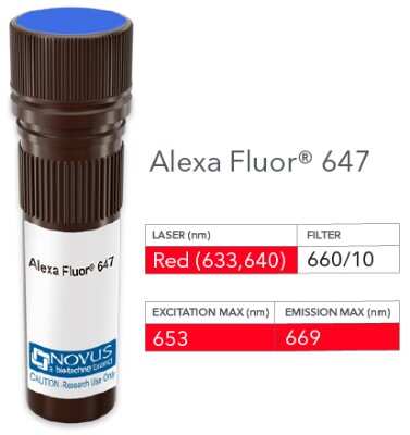
Product Image: HIF-1 alpha Antibody (H1alpha67) [Alexa Fluor® 647] [NB100-105AF647] - Vial of Alexa Fluor 647 conjugated antibody. Alexa Fluor 647 is optimally excited at 653 nm by the Red laser (633 or 640 nm) and has an emission maximum of 669 nm.
Applications for HIF-1 alpha Antibody (H1alpha67) [Alexa Fluor® 647]
Application
Recommended Usage
ELISA
Optimal dilutions of this antibody should be experimentally determined.
Flow Cytometry
Optimal dilutions of this antibody should be experimentally determined.
Immunoblotting
Optimal dilutions of this antibody should be experimentally determined.
Immunocytochemistry/Immunofluorescence
Optimal dilutions of this antibody should be experimentally determined.
Immunohistochemistry
Optimal dilutions of this antibody should be experimentally determined.
Immunohistochemistry-Frozen
Optimal dilutions of this antibody should be experimentally determined.
Immunohistochemistry-Paraffin
Optimal dilutions of this antibody should be experimentally determined.
Knockdown Validated
Optimal dilutions of this antibody should be experimentally determined.
Knockout Validated
Optimal dilutions of this antibody should be experimentally determined.
Product Image
Optimal dilutions of this antibody should be experimentally determined.
Western Blot
Optimal dilutions of this antibody should be experimentally determined.
Application Notes
Optimal dilution of this antibody should be experimentally determined.
Please Note: Optimal dilutions of this antibody should be experimentally determined.
Formulation, Preparation, and Storage
Purification
Protein G purified
Formulation
50mM Sodium Borate
Preservative
0.05% Sodium Azide
Concentration
Please see the vial label for concentration. If unlisted please contact technical services.
Shipping
The product is shipped with polar packs. Upon receipt, store it immediately at the temperature recommended below.
Stability & Storage
Store at 4C in the dark.
Background: HIF-1 alpha/HIF1A
HIF-1 or hypoxia inducible factor 1 (predicted molecular weight 93kDa), is a transcription factor commonly referred to as a "master regulator of the hypoxic response" for its central role in the regulation of cellular adaptations to hypoxia. In its active form under hypoxic conditions, HIF-1 is stabilized by the formation of a heterodimer of HIF-1 alpha and ARNT/HIF-1 beta subunits. Nuclear HIF-1 engages p300/CBP for binding to hypoxic response elements (HREs). This process induces transcription and regulation of genes including EPO, VEGF, iNOS2, ANGPT1 and OCT4 (4,5).
Under normoxic conditions, the HIF-1 alpha subunit is rapidly targeted and degraded by the ubiquitin proteasome system. This process is mediated by prolyl hydroxylase domain enzymes (PHDs), which catalyze the hydroxylation of key proline residues (Pro-402 and Pro-564) within the oxygen-dependent degradation domain of HIF-1 alpha. Once hydroxylated, HIF-1 alpha binds the von Hippel-Lindau tumor suppressor protein (pVHL) for subsequent ubiquitination and proteasomal degradation (4). pVHL dependent regulation of HIF-1 alpha plays a role in normal physiology and disease states. Regulation of HIF-1 alpha by pVHL is critical for the suppressive function of FoxP3+ regulatory Tcells (6). Repression of pVHL expression in chronic lymphocytic leukemia (CLL) B cells leads to HIF-1 alpha stabilization and increased VEGF secretion (7).
References
1. Semenza, G. L., Agani, F., Feldser, D., Iyer, N., Kotch, L., Laughner, E., & Yu, A. (2000). Hypoxia, HIF-1, and the pathophysiology of common human diseases. Advances in Experimental Medicine and Biology.
2. Muz, B., de la Puente, P., Azab, F., & Azab, A. K. (2015). The role of hypoxia in cancer progression, angiogenesis, metastasis, and resistance to therapy. Hypoxia. https://doi.org/10.2147/hp.s93413
3. Huang, Y., Lin, D., & Taniguchi, C. M. (2017). Hypoxia inducible factor (HIF) in the tumor microenvironment: friend or foe? Science China Life Sciences. https://doi.org/10.1007/s11427-017-9178-y
4. Koyasu, S., Kobayashi, M., Goto, Y., Hiraoka, M., & Harada, H. (2018). Regulatory mechanisms of hypoxia-inducible factor 1 activity: Two decades of knowledge. Cancer Science. https://doi.org/10.1111/cas.13483
5. Dengler, V. L., Galbraith, M. D., & Espinosa, J. M. (2014). Transcriptional regulation by hypoxia inducible factors. Critical Reviews in Biochemistry and Molecular Biology. https://doi.org/10.3109/10409238.2013.838205
6. Lee, J. H., Elly, C., Park, Y., & Liu, Y. C. (2015). E3Ubiquitin Ligase VHL Regulates Hypoxia-Inducible Factor-1 alpha to Maintain Regulatory T Cell Stability and Suppressive Capacity. Immunity. https://doi.org/10.1016/j.immuni.2015.05.016
7. Ghosh, A. K., Shanafelt, T. D., Cimmino, A., Taccioli, C., Volinia, S., Liu, C. G., ... Kay, N. E. (2009). Aberrant regulation of pVHL levels by microRNA promotes the HIF/VEGF axis in CLL B cells. Blood. https://doi.org/10.1182/blood-2008-10-185686
Long Name
Hypoxia Inducible Factor 1 Subunit Alpha
Alternate Names
BHLHE78, HIF 1A, HIF-1a, HIF1 alpha, HIF1A, MOP1, PASD8, H1alpha67
Gene Symbol
HIF1A
Additional HIF-1 alpha/HIF1A Products
Product Documents for HIF-1 alpha Antibody (H1alpha67) [Alexa Fluor® 647]
Product Specific Notices for HIF-1 alpha Antibody (H1alpha67) [Alexa Fluor® 647]
Alexa Fluor (R) products are provided under an intellectual property license from Life Technologies Corporation. The purchase of this product conveys to the buyer the non-transferable right to use the purchased product and components of the product only in research conducted by the buyer (whether the buyer is an academic or for-profit entity). The sale of this product is expressly conditioned on the buyer not using the product or its components, or any materials made using the product or its components, in any activity to generate revenue, which may include, but is not limited to use of the product or its components: (i) in manufacturing; (ii) to provide a service, information, or data in return for payment; (iii) for therapeutic, diagnostic or prophylactic purposes; or (iv) for resale, regardless of whether they are resold for use in research. For information on purchasing a license to this product for purposes other than as described above, contact Life Technologies Corporation, 5791 Van Allen Way, Carlsbad, CA 92008 USA or outlicensing@lifetech.com. This conjugate is made on demand. Actual recovery may vary from the stated volume of this product. The volume will be greater than or equal to the unit size stated on the datasheet.
This product is for research use only and is not approved for use in humans or in clinical diagnosis. Primary Antibodies are guaranteed for 1 year from date of receipt.
Loading...
Loading...
Loading...
Loading...
Loading...
Loading...