Human CD83 Fluorescein-conjugated Antibody
R&D Systems, part of Bio-Techne | Catalog # FAB1774F


Key Product Details
Species Reactivity
Validated:
Cited:
Applications
Validated:
Cited:
Label
Antibody Source
Product Specifications
Immunogen
Specificity
Clonality
Host
Isotype
Scientific Data Images for Human CD83 Fluorescein-conjugated Antibody
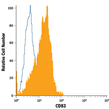
Detection of CD83 in Human CD14+Monocyte-derived Dendritic Cells by Flow Cytometry.
Human CD14+monocyte-derived dendritic cells were stained with Mouse Anti-Human CD83 Fluorescein-conjugated Monoclonal Antibody (Catalog # FAB1774F, filled histogram) or isotype control antibody (Catalog # IC002F, open histogram). View our protocol for Staining Membrane-associated Proteins.Applications for Human CD83 Fluorescein-conjugated Antibody
Flow Cytometry
Sample: Human CD14+ monocyte-derived dendritic cells
Formulation, Preparation, and Storage
Purification
Formulation
Shipping
Stability & Storage
- 12 months from date of receipt, 2 to 8 °C as supplied.
Background: CD83
Human CD83 is a 40‑50 kDa member of the Siglec (or sialic-acid-binding immunoglobulin-like lectin) family of transmembrane proteins (1, 2, 3). CD83 is synthesized as a type I transmembrane glycoprotein that contains a 125 amino acid (aa) extracellular region, a 22 aa transmembrane segment, and 39 aa cytoplasmic domain. It contains one V type Ig-like domain in the extracellular region with no inhibitory cytoplasmic motif(s). Although in vitro studies suggest CD83 may form membrane-bound covalent homodimers, in vivo this does not appear to be the case (1, 4). In the extracellular region, mouse and human CD83 are 66% aa identical (1, 2, 4, 5). Relative to human, mouse CD83 is 11 aa shorter in its extracellular domain and is expressed as a 30‑35 kDa protein (1, 4, 5). Human CD83 is active in the mouse system (4). One alternate splice form has been reported. This leads to a small monomeric soluble form of 74 aa that includes aa 20‑52 and aa 164‑205 (6, 7). In human, proteolytic cleavage and solubilization of CD83 has also been suggested, and this could lead to dimeric circulating CD83 (4, 6). CD83 is a primary marker for dendritic cells (3, 6, 8). It is also found on B cells (6, 9), neutrophils (10), monocytes and macrophages (11). Except for dendritic cells, CD83 expression is often transient. CD83 binds to sialic acids on target cells (12). Membrane CD83 appears to promote T cell proliferation, particularly of CD8+ cytotoxic T cells (13, 14). Soluble CD83, however, appears to be immunosuppressive and blocks T cell activation (15, 16). On monocytes, CD83 is suggested to drive monocytes into a fibrocyte phenotype (13). A lack of membrane-expressed CD83 leads to an unusual IL-4/IL-10 producing CD4+ T cell phenotype (17).
References
- Zhou, L-J. et al. (1992) J. Immunol. 149:735.
- Kozlow, E.J. et al. (1993) Blood 81:454.
- Fujimoto, Y and T.F. Tedder (2006) J. Med. Dent. Sci. 53:85.
- Lechmann, M. et al. (2005) Biochem. Biophys. Res. Commun. 329:132.
- Berchtold, S. et. al. (1999) FEBS Lett. 461:211.
- Hock, B.D. et al. (2001) Int. Immunol. 13:959.
- Dudziak, D. et al. (2005) J. Immunol. 174:6672.
- Velten, F.W. et al. (2007) Mol. Immunol. 44:1544.
- Cramer, S.O. et al. (2000) Int. Immunol. 12:1347.
- Yamashiro, S. et al. (2000) Blood 96:3958.
- Cao, W. et al. (2005) Biochem. J. 385:85.
- Scholler, N. et al. (2001) J. Immunol. 166:3865.
- Scholler, N. et al. (2002) J. Immunol. 168:2599.
- Hirano, N. et al. (2006) Blood 107:1528.
- Kotzor, N. et al. (2004) Immunobiology 209:129.
- Zinser, E. et al. (2006) Immunobiology 211:449.
- Garcia-Martinez, L.F. et al. (2004) J. Immunol. 173:2995.
Alternate Names
Gene Symbol
Additional CD83 Products
Product Documents for Human CD83 Fluorescein-conjugated Antibody
Product Specific Notices for Human CD83 Fluorescein-conjugated Antibody
For research use only