Human/Mouse ATG7 Antibody
R&D Systems, part of Bio-Techne | Catalog # MAB6608


Key Product Details
Validated by
Species Reactivity
Validated:
Cited:
Applications
Validated:
Cited:
Label
Antibody Source
Product Specifications
Specificity
Clonality
Host
Isotype
Scientific Data Images for Human/Mouse ATG7 Antibody
Detection of Human ATG7 by Western Blot.
Western blot shows lysates of HeLa human cervical epithelial carcinoma cell line and HepG2 human hepatocellular carcinoma cell line. PVDF membrane was probed with 2 µg/mL of Mouse Anti-Human ATG7 Monoclonal Antibody (Catalog # MAB6608) followed by HRP-conjugated Anti-Mouse IgG Secondary Antibody (Catalog # HAF007). A specific band was detected for ATG7 at approximately 75 kDa (as indicated). This experiment was conducted under reducing conditions and using Immunoblot Buffer Group 1.ATG7 in RAW 264.7 Mouse Cell Line.
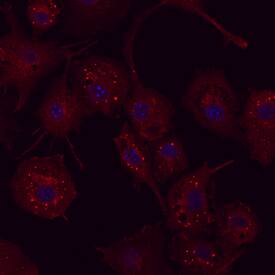
ATG7 was detected in immersion fixed RAW 264.7 mouse monocyte/macrophage cell line using Mouse Anti-Human ATG7 Monoclonal Antibody (Catalog # MAB6608) at 25 µg/mL for 3 hours at room temperature. Cells were stained using the Northern-Lights™ 557-conjugated Anti-Mouse IgG Secondary Antibody (red; Catalog # NL007) and counterstained with DAPI (blue). Specific staining was localized to autophagosomes. View our protocol for Fluorescent ICC Staining of Non-adherent Cells.ATG7 in Human Brain.
ATG7 was detected in formalin fixed paraffin-embedded sections of human brain (cortex) using Mouse Anti-Human ATG7 Monoclonal Antibody (Catalog # MAB6608) at 15 µg/mL overnight at 4 °C. Before incubation with the primary antibody, tissue was subjected to heat-induced epitope retrieval using Antigen Retrieval Reagent-Basic (Catalog # CTS013). Tissue was stained using the Anti-Mouse HRP-DAB Cell & Tissue Staining Kit (brown; Catalog # CTS002) and counter-stained with hematoxylin (blue). Specific staining was localized to neuronal cell bodies and processes. View our protocol for Chromogenic IHC Staining of Paraffin-embedded Tissue Sections.Applications for Human/Mouse ATG7 Antibody
CyTOF-ready
Immunocytochemistry
Sample: Immersion fixed RAW 264.7 mouse monocyte/macrophage cell line
Immunohistochemistry
Sample: Immersion fixed paraffin-embedded sections of human brain (cortex)
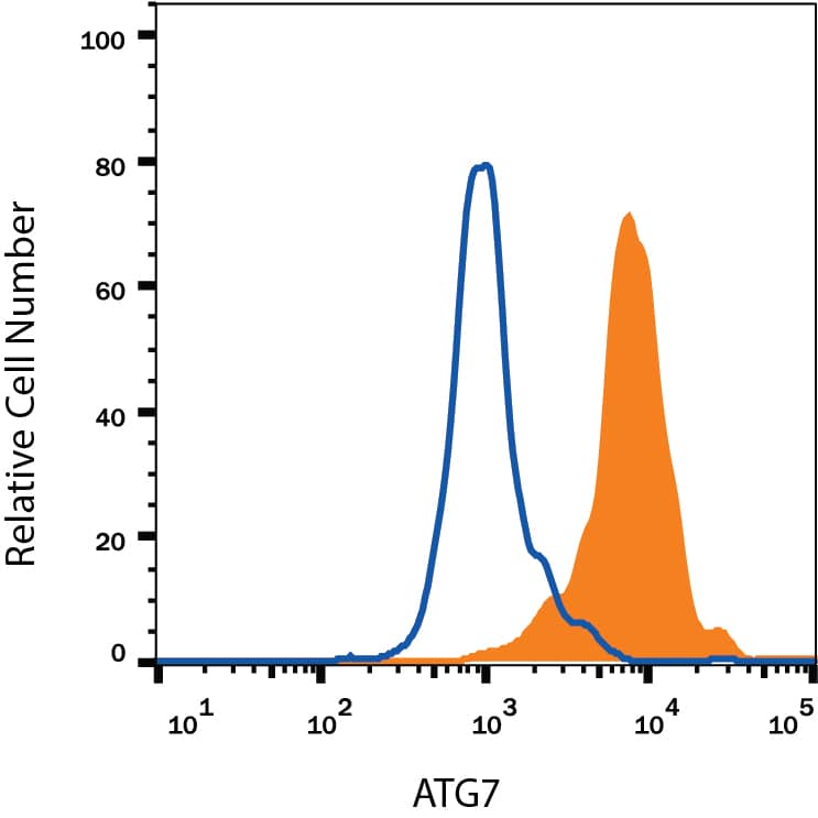
Intracellular Staining by Flow Cytometry
Sample: HeLa human cervical epithelial carcinoma cell line fixed with Flow Cytometry Fixation Buffer (Catalog # FC004) and permeabilized with Flow Cytometry Permeabilization/Wash Buffer I (Catalog # FC005)
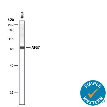
Simple Western
Sample: HeLa human cervical epithelial carcinoma cell line
Western Blot
Sample: HeLa human cervical epithelial carcinoma cell line and HepG2 human hepatocellular carcinoma cell line
Reviewed Applications
Read 3 reviews rated 5 using MAB6608 in the following applications:
Formulation, Preparation, and Storage
Reconstitution
Formulation
Shipping
Stability & Storage
Background: ATG7
ATG7, also known as APG7, is a 78 kDa cytosolic ubiquitin-E1-like enzyme that plays a key role in the autophagic pathway of intracellular bulk degradation. It is required for the conjugation of ATG5 to ATG12, the lipidation of ATG8, and subsequent autophagosome formation. ATG7 is required for mitochondrial removal during erythropoiesis and for the maintenance of axonal homeostasis. Alternate splicing of human ATG7 generates an isoform that lacks 31 aa at the C-terminus, a region that is required for ATG8 lipidation. Human ATG7 shares 93% aa sequence identitiy with mouse and rat ATG7.
Long Name
Alternate Names
Gene Symbol
UniProt
Additional ATG7 Products
Product Documents for Human/Mouse ATG7 Antibody
Product Specific Notices for Human/Mouse ATG7 Antibody
For research use only


