Human VEGFR3/Flt-4 Antibody
R&D Systems, part of Bio-Techne | Catalog # AF349


Key Product Details
Species Reactivity
Validated:
Cited:
Applications
Validated:
Cited:
Label
Antibody Source
Product Specifications
Immunogen
Tyr25-Ile776
Accession # P35916
Specificity
Clonality
Host
Isotype
Scientific Data Images for Human VEGFR3/Flt-4 Antibody
VEGFR3/Flt‑4 in Human Cervical Squamous Metaplasia.
VEGFR3/Flt-4 was detected in immersion fixed paraffin-embedded sections of human cervical squamous metaplasia using Human VEGFR3/Flt-4 Antigen Affinity-purified Polyclonal Antibody (Catalog # AF349) at 15 µg/mL overnight at 4 °C. Tissue was stained using the Anti-Goat HRP-DAB Cell & Tissue Staining Kit (brown; Catalog # CTS008) and counterstained with hematoxylin (blue). View our protocol for Chromogenic IHC Staining of Paraffin-embedded Tissue Sections.Detection of Human VEGFR3/Flt-4 by Western Blot
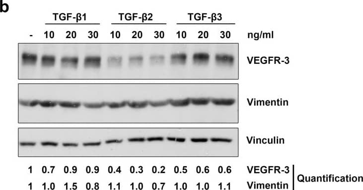
TGF-beta 1, -beta 2 and -beta 3 reduce lymphatic marker expression in LECs.Primary human LECs were treated with 10, 20 or 30 ng/ml TGF-beta 1, -beta 2 and -beta 3 for 72 hours (a) or 100 hours (b). Untreated cells served as a control. Lysates were prepared and analysed by Western blot using antibodies specific for Lyve-1, Prox-1, VEGFR-3 or vimentin. Vinculin served as loading control. The experiment was performed twice with equivalent results. For densitometry evaluation, protein bands were analysed using the software ImageJ. Bands for the Prox-1, Lyve-1, vimentin and VEGFR-3 proteins were normalized to the corresponding loading control and are displayed as the expression level relative to the untreated control samples. Image collected and cropped by CiteAb from the following open publication (https://dx.plos.org/10.1371/journal.pone.0162221), licensed under a CC-BY license. Not internally tested by R&D Systems.Detection of Human VEGFR3/Flt-4 by Immunocytochemistry/ Immunofluorescence
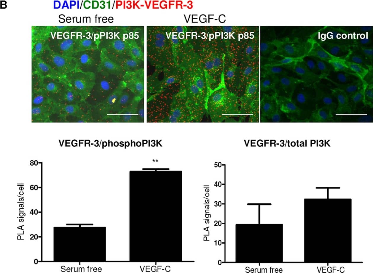
Direct interaction between PI3K p85 and VEGFR-3.A, LEC whole cell lysate was immunoprecipitated with anti-PI3K or isotype control IgG followed by Western blotting of phospho-tyrosine (p-Tyr), VEGFR-3 or PI3K. LECs were treated with vehicle control (serum-free media ‘0′), IgG control, VEGF-C (100 ng/ml, C) or VEGF-A (100 ng/ml, A) ligand for 15 minutes. Control untreated LEC whole cell lysate is indicate by ‘WCL’ and Jurkat cell lysate served as a positive control (Jurkat). B, Detection of VEGFR-3/phospho-PI3K complexes (red spots) in vitro in LECs grown in absence (B, upper left panel) or presence (B, upper middle panel) of VEGF-C (100 ng/ml) for 15 minutes; CD31 staining (green); DAPI (blue); isotype IgG control in PLA (B, upper right panel); quantification of VEGFR-3/phospho-PI3K and VEGFR-3/total-PI3K PLA signals using Olink Imaging Software (B, lower panel). Data shown as mean±s.e.m. **P0.01 using t-test analysis. n = 3. Bar = 50 µm. Image collected and cropped by CiteAb from the following open publication (https://dx.plos.org/10.1371/journal.pone.0039558), licensed under a CC-BY license. Not internally tested by R&D Systems.Applications for Human VEGFR3/Flt-4 Antibody
Immunohistochemistry
Sample: Immersion fixed paraffin-embedded sections of human lung and human cervical squamous metaplasia
Western Blot
Sample: Recombinant Human VEGFR3/Flt‑4 Fc Chimera (Catalog # 349-F4)
Formulation, Preparation, and Storage
Purification
Reconstitution
Formulation
Shipping
Stability & Storage
- 12 months from date of receipt, -20 to -70 °C as supplied.
- 1 month, 2 to 8 °C under sterile conditions after reconstitution.
- 6 months, -20 to -70 °C under sterile conditions after reconstitution.
Background: VEGFR3/Flt-4
VEGFR2 (KDR/Flk-1), VEGFR1 (Flt-1) and VEGFR3 (Flt-4) belong to the class III subfamily of receptor tyrosine kinases (RTKs). All three receptors contain seven immunoglobulin-like repeats in their extracellular domains and kinase insert domains in their intracellular regions. The expression of VEGFR1, 2, and 3 is almost exclusively restricted to the endothelial cells. These receptors are likely to play essential roles in vasculogenesis and angiogenesis.
VEGFR3 cDNA encodes a 1298 amino acid (aa) residue precursor protein with a 24 aa residue signal peptide. Mature VEGFR3 is composed of a 751 aa residue extracellular domain, a 22 aa residue transmembrane domain and a 482 aa residue cytoplasmic domain. Both VEGF-C and VEGF-D have been shown to bind and activate VEGFR3 (Flt-4). VEGFR3 is widely expressed in the early embryo but becomes restricted to lymphatic endothelia at later stages of development. It is likely that VEGFR3 may be important for lymph angiogenesis.
References
- Ferra, N. and R. Davis-Smyth (1997) Endocrine Reviews 18:4.
Long Name
Alternate Names
Gene Symbol
UniProt
Additional VEGFR3/Flt-4 Products
Product Documents for Human VEGFR3/Flt-4 Antibody
Product Specific Notices for Human VEGFR3/Flt-4 Antibody
For research use only


