LC3B Antibody (1251A) - Azide and BSA Free
Novus Biologicals, part of Bio-Techne | Catalog # NBP2-80829
Recombinant Monoclonal Antibody


Conjugate
Catalog #
Forumulation
Catalog #
Key Product Details
Species Reactivity
Validated:
Human, Mouse, Rat
Applications
CyTOF-ready, Flow (Intracellular), Flow Cytometry, Immunocytochemistry/Immunofluorescence, Immunohistochemistry, Immunohistochemistry-Paraffin, Immunoprecipitation, Knockout Validated, Western Blot
Label
Unconjugated
Antibody Source
Recombinant Monoclonal Rabbit IgG Clone # 1251A
Format
Azide and BSA Free
Concentration
1 mg/ml
Product Summary for LC3B Antibody (1251A) - Azide and BSA Free
Immunogen
Recombinant monoclonal LC3B Antibody (1251A) was made to a synthetic peptide made to an N-terminal portion of the human LC3B protein sequence (between residues 1-100). [UniProt# Q9GZQ8].
Reactivity Notes
Mouse and Rat reactivity reported from verified customer reviews.
Localization
Type I form of LC3B is cytoplasmic, whereas the type II form of LC3B binds to the autophagic membranes.
Clonality
Monoclonal
Host
Rabbit
Isotype
IgG
Theoretical MW
14.688 kDa.
Disclaimer note: The observed molecular weight of the protein may vary from the listed predicted molecular weight due to post translational modifications, post translation cleavages, relative charges, and other experimental factors.
Disclaimer note: The observed molecular weight of the protein may vary from the listed predicted molecular weight due to post translational modifications, post translation cleavages, relative charges, and other experimental factors.
Scientific Data Images for LC3B Antibody (1251A) - Azide and BSA Free
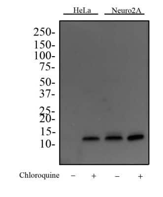
Western Blot: LC3B Antibody (1251A) - Azide and BSA Free [NBP2-80829] - Western Blot image of monoclonal anti-LC3B Antibody (Clone 1251D) [Catalog # NBP2-46892]. HeLa and Neuro2A cells were treated with or without 50 uM chloroquine for 24 hours as indicated. Whole cell protein was then separated on a 4-15% gel by SDS-PAGE, transferred to 0.2 um PVDF membrane for 30 min and blocked in 5% non-fat milk in TBST (Tris-buffered saline, 0.1% Tween 20). The membrane was probed with 2 ug/ml anti-LC3B Antibody in 1% milk, and detected with an anti-rabbit HRP secondary antibody using chemiluminescence. Note the accumulation of LC3B II upon chloroquine treatment. Bands for LC3 were detected at a molecular weight of approximately 15 kDa in treated HeLA cells, and both treated and untreated Neuro2A cells. Image from the standard format of this antibody.

Immunocytochemistry/Immunofluorescence: LC3B Antibody (1251A) - Azide and BSA Free [NBP2-80829] - HeLa cells were treated with Chlorquine for 24 hours prior to fixation, permeabilization and incubation with anti- [Catalog # NBP2-46892] and anti tubulin (NB100-690) antibodies. Image enlargement shows the accumulation of LC3 (green) on autophagosomes in response to chloroquine treatment. Tubulin staining is shown in red and DNA is counterstained with DAPI (blue). Image from the standard format of this antibody.

Immunohistochemistry-Paraffin: LC3B Antibody (1251A) - Azide and BSA Free [NBP2-80829] - IHC (Immunohistochemical) analysis of a formalin fixed and paraffin embedded tissue section of normal mouse brain using rabbit monoclonal LC3B (1251A) antibody [Catalog # NBP2-46892] at 1:100 dilution with HRP-DAB detection. The antibody generated a weak


Applications for LC3B Antibody (1251A) - Azide and BSA Free
Application
Recommended Usage
Flow (Intracellular)
1 - 2.5 ug/ml
Immunocytochemistry/Immunofluorescence
: 10-20 ug/ml
Immunohistochemistry
: 1:100-1:500
Immunoprecipitation
: 2-10 ug
Application Notes
In WB this LC3B recombinant monoclonal antibody detects both LC3B I and LC3B II with chloroquine treatment. With ICC autophagosome staining was observed after treatment with chloroquine.
Please Note: Optimal dilutions of this antibody should be experimentally determined.
Formulation, Preparation, and Storage
Purification
Protein G purified
Formulation
PBS
Format
Azide and BSA Free
Preservative
No Preservative
Concentration
1 mg/ml
Shipping
The product is shipped with polar packs. Upon receipt, store it immediately at the temperature recommended below.
Stability & Storage
Store at 4C short term. Aliquot and store at -20C long term. Avoid freeze-thaw cycles.
Background: LC3B
Autophagic flux is supported by autophagy-related proteins (Atgs) initially identified in yeast (6,7). The core autophagy machinery is comprised of 17 Atg proteins that play specific roles in autophagosome formation. Among these Atg proteins, Atg8 is not only involved in autophagosome formation but also functions in cargo selection. In mammals, several Atg8 homologues have been identified including microtubule-associated protein 1 light chain 3 alpha, beta and gamma - LC3A, LC3B, and LC3C (8) respectively, as well as GABA type A receptor-associated protein (GABARAP), GABARAP-Like1, and GABARAP-Like2 (9). LC3 (predicted molecular weight 14kD) is ubiquitously expressed and undergoes posttranslational processing after synthesis. First, the cysteine protease Atg4 cleaves a carboxy terminal sequence to generate the cytosolic form LC3-I. Next, E1-like (Atg7) and E2-like (Atg3) enzymes conjugate phosphatidylethanolamine to the newly exposed carboxyterminal glycine, generating LC3-II. Finally, the Atg12-Atg5-Atg16L1 complex participates in LC3 lipidation and autophagosome formation (10). LC3B-I to LC3B-II conversion correlates with autophagosome number and is considered the best marker to monitor autophagy.
References
1. Yu, L., Chen, Y., & Tooze, S. A. (2018). Autophagy pathway: Cellular and molecular mechanisms. Autophagy. https://doi.org/10.1080/15548627.2017.1378838
2. Forrester, A., De Leonibus, C., Grumati, P., Fasana, E., Piemontese, M., Staiano, L., ... Settembre, C. (2019). A selective ER -phagy exerts procollagen quality control via a Calnexin- FAM 134B complex. The EMBO Journal. https://doi.org/10.15252/embj.201899847
3. He, X., Zhu, Y., Zhang, Y., Geng, Y., Gong, J., Geng, J., ... Zhong, H. (2019). RNF34 functions in immunity and selective mitophagy by targeting MAVS for autophagic degradation. The EMBO Journal. https://doi.org/10.15252/embj.2018100978
4. Mathai, B., Meijer, A., & Simonsen, A. (2017). Studying Autophagy in Zebrafish. Cells. https://doi.org/10.3390/cells6030021
5. Losier, T. T., Akuma, M., McKee-Muir, O. C., LeBlond, N. D., Suk, Y., Alsaadi, R. M., ... Russell, R. C. (2019). AMPK Promotes Xenophagy through Priming of Autophagic Kinases upon Detection of Bacterial Outer Membrane Vesicles. Cell Reports. https://doi.org/10.1016/j.celrep.2019.01.062
6. Nakatogawa, H., Suzuki, K., Kamada, Y., & Ohsumi, Y. (2009). Dynamics and diversity in autophagy mechanisms: Lessons from yeast. Nature Reviews Molecular Cell Biology. https://doi.org/10.1038/nrm2708
7. Tsukada, M., & Ohsumi, Y. (1993). Isolation and characterization of autophagy-defective mutants of Saccharomyces cerevisiae. FEBS Letters. https://doi.org/10.1016/0014-5793(93)80398-E
8. Wild, P., McEwan, D. G., & Dikic, I. (2014). The LC3 interactome at a glance. Journal of Cell Science. https://doi.org/10.1242/jcs.140426
9. Igloi, G. L. (2001). Cloning, expression patterns, and chromosome localization of three human and two mouse homologues of GABAA receptor-associated protein. Genomics. https://doi.org/10.1006/geno.2001.6555
10. Glick, D., Barth, S., & Macleod, K. F. (2010). Autophagy: Cellular and molecular mechanisms. Journal of Pathology. https://doi.org/10.1002/path.2697
Long Name
Microtubule-associated Protein 1 Light Chain 3 beta
Alternate Names
Apg8b, ATG8F, LC3II, MAP1LC3B
Gene Symbol
MAP1LC3B
Additional LC3B Products
Product Documents for LC3B Antibody (1251A) - Azide and BSA Free
Product Specific Notices for LC3B Antibody (1251A) - Azide and BSA Free
This product is for research use only and is not approved for use in humans or in clinical diagnosis. Primary Antibodies are guaranteed for 1 year from date of receipt.
Loading...
Loading...
Loading...
Loading...
Loading...