LC3B Antibody [PE]
Novus Biologicals, part of Bio-Techne | Catalog # NB600-1384PE

Conjugate
Catalog #
Forumulation
Catalog #
Key Product Details
Species Reactivity
Validated:
Human, Mouse, Rat, Porcine, Bacteria, Bovine, Canine, Primate, Yeast, Zebrafish
Applications
Flow Cytometry, Knockdown Validated, Knockout Validated, Product Image
Label
PE (Excitation = 488 nm, Emission = 575 nm)
Antibody Source
Polyclonal Rabbit IgG
Concentration
Please see the vial label for concentration. If unlisted please contact technical services.
Product Summary for LC3B Antibody [PE]
Immunogen
Polyclonal LC3B Antibody was made to a synthetic peptide made to the N-terminal region of the human LC3B protein.
Reactivity Notes
Use in Yeast reported in scientific literature (PMID:35247568).Use in Rat reported in scientific literature (PMID:34499623). Mouse reactivity reported in scientific literature (PMID:32802192). Zebrafish reactivity reported in scientific literature (PMID: 23724125). . Canine and primate reactivity reported in scientific literature (PMID: 24027311). Porcine reactivity reported in scientific literature (PMID: 25378587). . Rat reactivity reported in scientific literature (30067379). . Bacteria reactivity reported in scientific literature (31110360). . Bovine reactivity reported in scientific literature (21868124). . Other species have not been tested.
Localization
LC3-I is cytoplasmic. LC3-II binds to the autophagic membranes.
Marker
Autophagosome Marker
Clonality
Polyclonal
Host
Rabbit
Isotype
IgG
Theoretical MW
14.688 kDa.
Disclaimer note: The observed molecular weight of the protein may vary from the listed predicted molecular weight due to post translational modifications, post translation cleavages, relative charges, and other experimental factors.
Disclaimer note: The observed molecular weight of the protein may vary from the listed predicted molecular weight due to post translational modifications, post translation cleavages, relative charges, and other experimental factors.
Scientific Data Images for LC3B Antibody [PE]
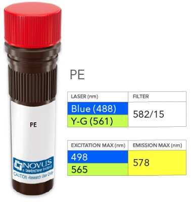
Product Image: LC3B Antibody [PE] [NB600-1384PE] - Vial of PE conjugated antibody. PE has two excitation maxima, 498 nm excited by the Blue laser (488 nm) and 565 nm excited by the Yellow-Green laser (561 nm). Both result in emission at 578 nm.
Applications for LC3B Antibody [PE]
Application
Recommended Usage
Flow Cytometry
Optimal dilutions of this antibody should be experimentally determined.
Knockdown Validated
Optimal dilutions of this antibody should be experimentally determined.
Knockout Validated
Optimal dilutions of this antibody should be experimentally determined.
Product Image
Optimal dilutions of this antibody should be experimentally determined.
Application Notes
Optimal dilution of this antibody should be experimentally determined.
Please Note: Optimal dilutions of this antibody should be experimentally determined.
Formulation, Preparation, and Storage
Purification
Immunogen affinity purified
Formulation
PBS
Preservative
0.05% Sodium Azide
Concentration
Please see the vial label for concentration. If unlisted please contact technical services.
Shipping
The product is shipped with polar packs. Upon receipt, store it immediately at the temperature recommended below.
Stability & Storage
Store at 4C in the dark.
Background: LC3B
Autophagic flux is supported by autophagy-related proteins (Atgs) initially identified in yeast (6,7). The core autophagy machinery is comprised of 17 Atg proteins that play specific roles in autophagosome formation. Among these Atg proteins, Atg8 is not only involved in autophagosome formation but also functions in cargo selection. In mammals, several Atg8 homologues have been identified including microtubule-associated protein 1 light chain 3 alpha, beta and gamma - LC3A, LC3B, and LC3C (8) respectively, as well as GABA type A receptor-associated protein (GABARAP), GABARAP-Like1, and GABARAP-Like2 (9). LC3 (predicted molecular weight 14kD) is ubiquitously expressed and undergoes posttranslational processing after synthesis. First, the cysteine protease Atg4 cleaves a carboxy terminal sequence to generate the cytosolic form LC3-I. Next, E1-like (Atg7) and E2-like (Atg3) enzymes conjugate phosphatidylethanolamine to the newly exposed carboxyterminal glycine, generating LC3-II. Finally, the Atg12-Atg5-Atg16L1 complex participates in LC3 lipidation and autophagosome formation (10). LC3B-I to LC3B-II conversion correlates with autophagosome number and is considered the best marker to monitor autophagy.
References
1. Yu, L., Chen, Y., & Tooze, S. A. (2018). Autophagy pathway: Cellular and molecular mechanisms. Autophagy. https://doi.org/10.1080/15548627.2017.1378838
2. Forrester, A., De Leonibus, C., Grumati, P., Fasana, E., Piemontese, M., Staiano, L., ... Settembre, C. (2019). A selective ER -phagy exerts procollagen quality control via a Calnexin- FAM 134B complex. The EMBO Journal. https://doi.org/10.15252/embj.201899847
3. He, X., Zhu, Y., Zhang, Y., Geng, Y., Gong, J., Geng, J., ... Zhong, H. (2019). RNF34 functions in immunity and selective mitophagy by targeting MAVS for autophagic degradation. The EMBO Journal. https://doi.org/10.15252/embj.2018100978
4. Mathai, B., Meijer, A., & Simonsen, A. (2017). Studying Autophagy in Zebrafish. Cells. https://doi.org/10.3390/cells6030021
5. Losier, T. T., Akuma, M., McKee-Muir, O. C., LeBlond, N. D., Suk, Y., Alsaadi, R. M., ... Russell, R. C. (2019). AMPK Promotes Xenophagy through Priming of Autophagic Kinases upon Detection of Bacterial Outer Membrane Vesicles. Cell Reports. https://doi.org/10.1016/j.celrep.2019.01.062
6. Nakatogawa, H., Suzuki, K., Kamada, Y., & Ohsumi, Y. (2009). Dynamics and diversity in autophagy mechanisms: Lessons from yeast. Nature Reviews Molecular Cell Biology. https://doi.org/10.1038/nrm2708
7. Tsukada, M., & Ohsumi, Y. (1993). Isolation and characterization of autophagy-defective mutants of Saccharomyces cerevisiae. FEBS Letters. https://doi.org/10.1016/0014-5793(93)80398-E
8. Wild, P., McEwan, D. G., & Dikic, I. (2014). The LC3 interactome at a glance. Journal of Cell Science. https://doi.org/10.1242/jcs.140426
9. Igloi, G. L. (2001). Cloning, expression patterns, and chromosome localization of three human and two mouse homologues of GABAA receptor-associated protein. Genomics. https://doi.org/10.1006/geno.2001.6555
10. Glick, D., Barth, S., & Macleod, K. F. (2010). Autophagy: Cellular and molecular mechanisms. Journal of Pathology. https://doi.org/10.1002/path.2697
Long Name
Microtubule-associated Protein 1 Light Chain 3 beta
Alternate Names
Apg8b, ATG8F, LC3II, MAP1LC3B
Gene Symbol
MAP1LC3B
Additional LC3B Products
Product Documents for LC3B Antibody [PE]
Product Specific Notices for LC3B Antibody [PE]
This product is for research use only and is not approved for use in humans or in clinical diagnosis. Primary Antibodies are guaranteed for 1 year from date of receipt.
Loading...
Loading...
Loading...
Loading...
Loading...
Loading...