MUC1 Antibody (SM3) - BSA Free
Novus Biologicals, part of Bio-Techne | Catalog # NB120-22711


Conjugate
Catalog #
Forumulation
Catalog #
Key Product Details
Species Reactivity
Validated:
Human, Mouse
Cited:
Human, Mouse
Applications
Validated:
ELISA, Flow Cytometry, Immunocytochemistry/Immunofluorescence, Immunohistochemistry, Immunohistochemistry-Frozen, Immunohistochemistry-Paraffin
Cited:
ELISA, Flow Cytometry, IF/IHC, Immunohistochemistry
Label
Unconjugated
Antibody Source
Monoclonal Mouse IgG2a Kappa Clone # SM3
Format
BSA Free
Concentration
1.0 mg/ml
Product Summary for MUC1 Antibody (SM3) - BSA Free
Immunogen
Hydrogen fluoride deglycosylated milk mucin.
Reactivity Notes
Mouse reactivity reported in scientific literature (PMID: 1372533) No record of testing in other species.
Localization
Cell Membrane and Cytoplasmic
Specificity
Reacts very little with normal tissue. Found to react with breast, colon and ovarian carcinomas and adinocarcinoma. SM3 recognises the under-glycosylated form of MUC1 and is therefore tumour specific.
Clonality
Monoclonal
Host
Mouse
Isotype
IgG2a Kappa
Scientific Data Images for MUC1 Antibody (SM3) - BSA Free
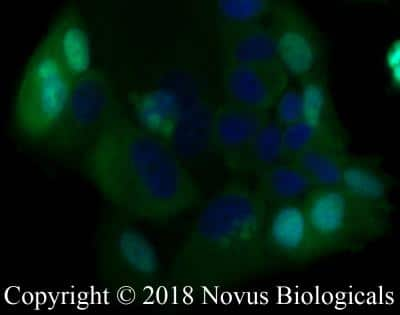
Immunocytochemistry/Immunofluorescence: MUC-1 Antibody (SM3) [NB120-22711] - MCF7 cells were fixed for 10 minutes using 10% formalin and then permeabilized for 5 minutes using 1X PBS + 0.05% Triton-X100. The cells were incubated with anti-MUC1 [SM3] at 5 ug/ml overnight at 4C and detected with an anti-mouse IgG Dylight 488 (Green) at a 1:500 dilution. Nuclei were counterstained with DAPI (Blue). Cells were imaged using a 40X objective.

Immunohistochemistry-Paraffin: MUC-1 Antibody (SM3) [NB120-22711] - IHC analysis of formalin fixed paraffin embedded tissue section of human breast cancer xenograft using MUC-1 antibody clone SM3 at 1:10 dilution. The xenograft section depicted a very specific and intense signal in the periphery of the cancer cells. The necrotic cells also developed a strong immune-positivity while the tumor stroma as well as the nuclei of cells were negative for immunostaining.

Flow Cytometry: MUC1 Antibody (SM3) - BSA Free [NB120-22711] - An intracellular stain was performed on MCF7 cells with MUC1 Antibody (SM3) NB120-22711AF700 (blue) and a matched isotype control (orange). Cells were fixed with 4% PFA and then permeabilized with 0.1% saponin. Cells were incubated in an antibody dilution of 5 ug/mL for 30 minutes at room temperature. Both antibodies were conjugated to Alexa Fluor 700.
Applications for MUC1 Antibody (SM3) - BSA Free
Application
Recommended Usage
ELISA
reported in multiple pieces of scientific literature
Flow Cytometry
reported in multiple pieces of scientific literature
Immunocytochemistry/Immunofluorescence
10 ug/ml
Immunohistochemistry
1:10-1:500
Immunohistochemistry-Frozen
1:10-1:500
Immunohistochemistry-Paraffin
1:10-1:500
Application Notes
IHC-Fr and IHC-P: Use at an assay dependent dilution.
Please Note: Optimal dilutions of this antibody should be experimentally determined.
Formulation, Preparation, and Storage
Purification
Protein G purified
Formulation
PBS
Format
BSA Free
Preservative
0.02% Sodium Azide
Concentration
1.0 mg/ml
Shipping
The product is shipped with polar packs. Upon receipt, store it immediately at the temperature recommended below.
Stability & Storage
Store at 4C short term. Aliquot and store at -20C long term. Avoid freeze-thaw cycles.
Background: MUC-1
Overexpression of mucins, including MUC1, is a feature of many epithelial cancers (1,3,5,6). The presence of truncated glycan structures called tumor-associated carbohydrate antigens (TACAs) on MUC1 play a role in cancer progression and a loss of apical-basal polarity (5). Carbohydrate-binding partners called lectins are the primary binding partners of TACAs that give rise to the pro-tumor microenvironment and metastasis (5). Given this unique feature, TACAs are a potential target for cancer immunotherapies (5). There are a number of vaccines, drugs, and antibodies targeting MUC1 for treatment of a variety of cancers including breast, lung, and prostate (6). In addition to a role in cancer progression, MUC1, and specifically the CT portion, has been shown to have a positive, anti-inflammatory role in a variety of lung and airway infections (7).
References
1. Khodabakhsh, F., Merikhian, P., Eisavand, M. R., & Farahmand, L. (2021). Crosstalk between MUC1 and VEGF in angiogenesis and metastasis: a review highlighting roles of the MUC1 with an emphasis on metastatic and angiogenic signaling. Cancer cell international. https://doi.org/10.1186/s12935-021-01899-8
2. Nath, S., & Mukherjee, P. (2014). MUC1: a multifaceted oncoprotein with a key role in cancer progression. Trends in molecular medicine. https://doi.org/10.1016/j.molmed.2014.02.007
3. Dhar, P., & McAuley, J. (2019). The Role of the Cell Surface Mucin MUC1 as a Barrier to Infection and Regulator of Inflammation. Frontiers in cellular and infection microbiology. https://doi.org/10.3389/fcimb.2019.00117
4. Uniprot (P15941)
5. Beckwith, D. M., & Cudic, M. (2020). Tumor-associated O-glycans of MUC1: Carriers of the glyco-code and targets for cancer vaccine design. Seminars in immunology. https://doi.org/10.1016/j.smim.2020.101389
6. Almasmoum H. (2021). The Roles of Transmembrane Mucins Located on Chromosome 7q22.1 in Colorectal Cancer. Cancer management and research. https://doi.org/10.2147/CMAR.S299089
7. Ballester, B., Milara, J., & Cortijo, J. (2021). The role of mucin 1 in respiratory diseases. European respiratory review : an official journal of the European Respiratory Society. https://doi.org/10.1183/16000617.0149-2020
Long Name
Mucin 1, Cell Surface-associated
Alternate Names
CD227, Episialin, H23AG, KL-6, Mucin-1, PEM, PEMT
Entrez Gene IDs
4582 (Human)
Gene Symbol
MUC1
UniProt
Additional MUC-1 Products
Product Documents for MUC1 Antibody (SM3) - BSA Free
Product Specific Notices for MUC1 Antibody (SM3) - BSA Free
This product is for research use only and is not approved for use in humans or in clinical diagnosis. Primary Antibodies are guaranteed for 1 year from date of receipt.
Loading...
Loading...
Loading...
Loading...
Loading...
Loading...