SDHB Antibody
Novus Biologicals, part of Bio-Techne | Catalog # NB600-793


Key Product Details
Species Reactivity
Validated:
Human
Applications
Control, Flow Cytometry, Immunocytochemistry/Immunofluorescence, Immunohistochemistry, Immunohistochemistry-Paraffin, Peptide ELISA, Western Blot
Label
Unconjugated
Antibody Source
Polyclonal Goat IgG
Concentration
0.5 mg/ml
Product Summary for SDHB Antibody
Immunogen
Peptide with sequence C-ATYKEKKASV corresponding to C-Terminus according to NP_002991.2.
Marker
Mitochondria Marker
Clonality
Polyclonal
Host
Goat
Isotype
IgG
Scientific Data Images for SDHB Antibody
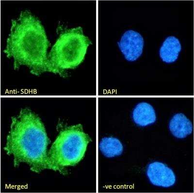
Immunocytochemistry/Immunofluorescence: SDHB Antibody [NB600-793] - Immunofluorescence analysis of paraformaldehyde fixed A431 cells, permeabilized with 0.15% Triton. Primary incubation 1hr (10ug/ml) followed by Alexa Fluor 488 secondary antibody (2ug/ml), showing plasma membrane and cytoplasmic staining. The nuclear stain is DAPI (blue). Negative control: Unimmunized goat IgG (10ug/ml) followed by Alexa Fluor 488 secondary antibody (2ug/ml).

Immunohistochemistry: SDHB Antibody [NB600-793] - (6ug/ml) staining of paraffin embedded Human Kidney. Heat induced antigen retrieval with citrate buffer pH 6, HRP-staining.

Flow Cytometry: SDHB Antibody [NB600-793] - Flow cytometric analysis of paraformaldehyde fixed A431 cells (blue line), permeabilized with 0.5% Triton. Primary incubation 1hr (10ug/ml) followed by Alexa Fluor 488 secondary antibody (1ug/ml). IgG control: Unimmunized goat IgG (black line) followed by Alexa Fluor 488 secondary antibody.
Applications for SDHB Antibody
Application
Recommended Usage
Flow Cytometry
10 ug/ml
Immunocytochemistry/Immunofluorescence
10 ug/ml
Immunohistochemistry
6.0 - 8.0 ug/ml
Immunohistochemistry-Paraffin
6.0 - 8.0 ug/ml
Peptide ELISA
Detection limit 1:8000
Western Blot
0.1 - 0.3 ug/ml
Application Notes
Western blot: Approx 28-30kDa band observed in lysates of cell lines HEK293, Jurkat and HeLa (calculated MW of 31.6kDa according to NP_002991.2). Recommended concentration: 0.1-0.3ug/ml. Primary incubation 1 hour at room temperature.
Please Note: Optimal dilutions of this antibody should be experimentally determined.
Formulation, Preparation, and Storage
Purification
Immunogen affinity purified
Formulation
Tris saline (20 mM Tris pH 7.3, 150 mM NaCl), 0.5% BSA
Preservative
0.02% Sodium Azide
Concentration
0.5 mg/ml
Shipping
The product is shipped with polar packs. Upon receipt, store it immediately at the temperature recommended below.
Stability & Storage
Store at -20C. Avoid freeze-thaw cycles.
Background: SDHB
Alternate Names
EC 1.3.5.1, FLJ92337, Ip, Iron-sulfur subunit of complex II, PGL4, SDH, SDH1, SDH2, SDHIP, succinate dehydrogenase [ubiquinone] iron-sulfur subunit, mitochondrial, succinate dehydrogenase complex, subunit B, iron sulfur (Ip)
Entrez Gene IDs
6390 (Human)
Gene Symbol
SDHB
UniProt
Additional SDHB Products
Product Documents for SDHB Antibody
Product Specific Notices for SDHB Antibody
This product is for research use only and is not approved for use in humans or in clinical diagnosis. Primary Antibodies are guaranteed for 1 year from date of receipt.
Loading...
Loading...
Loading...
Loading...
Loading...