Mouse G-CSF ELISA Kit - Quantikine

R&D Systems, part of Bio-Techne | Catalog # MCS00

R&D Systems, part of Bio-Techne
Catalog #
Availability
Size / Price
Qty
Loading...
MCS00

Key Product Details

Assay Length

4.5 hours

Sample Type & Volume Required Per Well

Cell Culture Supernates (25 µL), Serum (50 µL)

Sensitivity

5 pg/mL

Assay Range

14.1-900 pg/mL (Cell Culture Supernates, Serum)

Product Summary for Mouse G-CSF Quantikine ELISA Kit

The Quantikine Mouse G-CSF Immunoassay is a 4.5 hour solid phase ELISA designed to measure mouse G-CSF in cell culture supernates and serum. It contains E. coli-expressed recombinant mouse G-CSF and antibodies raised against the recombinant factor. This immunoassay has been shown to quantitate the recombinant mouse G-CSF accurately. Results obtained using natural mouse G-CSF showed dose-response curves that were parallel to the standard curves obtained using the recombinant Quantikine kit standards. These results indicate that this kit can be used to determine relative mass values for natural mouse G-CSF.

Product Specifications

Assay Type

Solid Phase Sandwich ELISA

Format

96-well strip plate

Measurement

Quantitative ELISA

Detection Method

Colorimetric - 450nm (TMB)

Conjugate

HRP

Reactivity

Mouse

Specificity

Natural and recombinant mouse G-CSF

Cross-reactivity

0.5% cross-reactivity observed with available related molecules. 50% cross-species reactivity observed with species tested.

Interference

No significant interference observed with available related molecules.

Precision

Intra-Assay Precision (Precision within an assay) Three samples of known concentration were tested on one plate to assess intra-assay precision.

Inter-Assay Precision (Precision between assays) Three samples of known concentration were tested in separate assays to assess inter-assay precision.

Cell Culture Supernates, Serum

Intra-Assay Precision Inter-Assay Precision
Sample 1 2 3 1 2 3
n 20 20 20 20 20 20
Mean (pg/mL) 64.4 196 697 64.8 195 620
Standard Deviation 2.3 8.8 43 4.1 15.5 50.6
CV% 3.6 4.5 6.2 6.3 7.9 8.2

Recovery for Mouse G-CSF Quantikine ELISA Kit

The recovery of mouse G-CSF spiked to three levels throughout the range of the assay in various matrices was evaluated.

Sample Type Average % Recovery Range %
Cell Culture Supernates (n=7) 92 82-110
Serum (n=5) 96 80-119

Linearity

To assess the linearity of the assay, five or more samples containing and/or spiked with various concentrations of mouse G-CSF in each matrix were diluted with Calibrator Diluent and then assayed.

Mouse G-CSF ELISA Linearity

Scientific Data Images for Mouse G-CSF Quantikine ELISA Kit

Mouse G-CSF ELISA Standard Curve

Mouse G-CSF ELISA Standard Curve

Detection of Mouse G-CSF by ELISA

Detection of Mouse G-CSF by ELISA

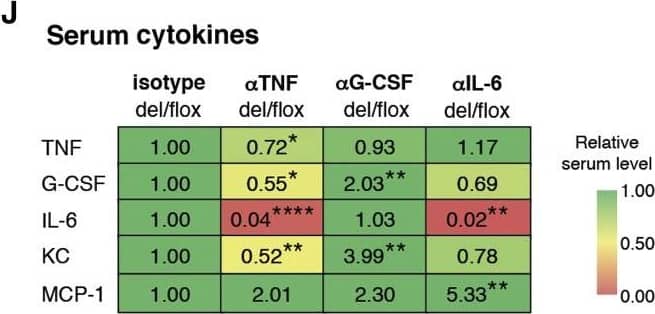
Neutralization of TNF Ameliorates Inflammation Caused by OTULIN Deficiency(A–J) Measurements from tamoxifen (tx)-treated (arrows) CreERT2-Otulinflox bone marrow chimeras injected with (A–C) anti-TNF neutralizing antibodies ( alphaTNF) (data were pooled from two independent experiments), (D–F) anti-G-CSF-neutralizing antibodies ( alphaG-CSF), (G–I) anti-IL-6-neutralizing antibodies ( alphaIL-6), or isotype control as indicated.(A, D, and G) Body weight of CreERT2-Otulinflox chimeric mice treated with neutralizing antibodies as indicated.(B, E, and H) Blood neutrophil counts from CreERT2-Otulinflox chimeric mice treated as indicated.(C, F, and I) Total number of infiltrating CD11b+Gr-1+ neutrophils in spleen and peritoneal lavage (PL) measured by flow cytometry from CreERT2-Otulinflox chimeric mice treated as indicated.(J) Heatmap of Luminex multiplex analysis of cytokines and chemokines in serum from terminal bleeds on days 6 or 7 from chimeric mice treated as indicated. Numbers indicate relative change compared to isotype-treated del/flox mice within each experiment. G-CSF levels for alphaG-CSF and alphaIL-6 were measured by ELISA. Asterisks (∗) indicate the level of statistical significance.(K) Model of TNF-driven systemic inflammation and the contributions from different cytokines in OTULIN-deficient mice.(A–J) Data are presented as mean ± SEM, and n represents number of mice.See also Figure S3. Image collected and cropped by CiteAb from the following publication (https://pubmed.ncbi.nlm.nih.gov/27523608), licensed under a CC-BY license. Not internally tested by R&D Systems.

Preparation and Storage

Shipping

The product is shipped with polar packs. Upon receipt, store it immediately at the temperature recommended below.

Stability & Storage

Store the unopened product at 2 - 8 °C. Do not use past expiration date.

Background: G-CSF

Mouse granulocyte-colony stimulating factor (G-CSF) is a 24-25 kDa monomeric glycoprotein that regulates the proliferation, differentiation and activation of hematopoietic cells. Mouse G-CSF cDNA encodes a 208 amino acid (aa) precursor protein with a 30 aa signal sequence that is proteolytically cleaved to form a 178 aa O-glycosylated mature protein containing two intrachain disulfide bridges. In humans, two distinct cDNA clones, encoding a 204 aa form and a minor alternatively spliced 207 aa form of G-CSF precursors, have been isolated. Mouse and human G-CSF are 76% identical at the aa sequence level and the two proteins show species cross-reactivity. G-CSF is produced primarily by monocytes and macrophages upon activation by endotoxin, TNF-alpha or IL-1. Other cell types, including fibroblasts, endothelial cells, astrocytes and bone marrow stroma cells, can also secrete G-CSF after activation. In addition, various tumor cells express G-CSF constitutively.  

Mouse G-CSF receptor (G-CSF R) is a 120 kDa, type I transmembrane glycoprotein that belongs to the hematopoietin receptor superfamily. The mature protein is 812 amino acids in length and contains a 601 aa extracellular region, a 24 aa transmembrane segment, and a 187 aa cytoplasmic domain. The extracellular region contains multiple modules, including an N-terminal Ig-like domain, a cytokine receptor homology domain, and three fibronectin type III domains. Based on crystallographic study, G-CSF receptor forms a complex with the ligand in a 2:2 ratio. Mouse and human G-CSF receptor share 63% aa sequence identity. Cells known to express G-CSF R include monocytes and neutrophils, megakaryocytes and platelets, CD34+ CD33+ and CD34+ CD38+ hematopoietic progenitors, trophoblasts, endothelial cells and various tumor cell types.  

G-CSF is an important regulator for granulopoiesis in vivo. It has been demonstrated that G-CSF can support the growth of multi-lineage hematopoietic progenitor cells without influencing their commitment to the myeloid lineage and mobilize hematopoietic progenitor cells from the bone marrow into the bloodstream. On mature neutrophils, G-CSF may regulate neutrophil survival by controlling their rate of apoptosis. G-CSF has also been shown to enhance the functional capacity of mature neutrophils. As a consequence of its effects on hematopoietic progenitor cells, G-CSF has been shown to enhance monocytopoiesis in the presence M-CSF. Within the peripheral blood stem cell population mobilized with G-CSF, selective increases in the number of T helper 2-inducing dendritic cells are found.

Long Name

Granulocyte Colony Stimulating Factor

Alternate Names

C17orf33, CSF3, CSF3OS, Filgrastim, GCSF, Lenograstim, Pluripoietin

Entrez Gene IDs

1440 (Human); 12985 (Mouse)

Gene Symbol

CSF3

Additional G-CSF Products

Product Documents for Mouse G-CSF Quantikine ELISA Kit

Certificate of Analysis

To download a Certificate of Analysis, please enter a lot number in the search box below.

Note: Certificate of Analysis not available for kit components.

Product Specific Notices for Mouse G-CSF Quantikine ELISA Kit

For research use only

Loading...
Loading...
Loading...

⚠ WARNING: This product can expose you to chemicals including N,N-Dimethylforamide, which is known to the State of California to cause cancer. For more information, go to www.P65Warnings.ca.gov.