Simple Western Charge

Charge-Based Separation Assays
The Simple Western Charge assay is an automated assay that doesn’t require the use of gels, transfer devices, blots, film or manual analysis where proteins are separated by their isoelectric point (pI) rather than their molecular weight. Simple Western Charge assays (using capillary isoelectric focusing, or cIEF) provide enhanced sensitivity and exquisite resolution for nearly all variant forms of a protein. Because proteins are separated by their pIs, you'll be able to see extremely small isoelectric point differences, to resolve post translational modifications such as phosphorylation and glycosylation that changes a proteins pI. It's a new way of looking at discrete changes in very small samples that you might just be surprised by!
How Simple Western Charge Works
Simple Western Charge assays take place in a capillary. Samples prepared in ampholytes and required reagents are loaded into an assay plate and placed in Peggy Sue™ or NanoPro™ 1000. The prepared sample is automatically loaded into the capillary and separated by charge according to their pI before the resolve proteins are immobilized to the capillary wall via a proprietary, photoactivated capture chemistry. Target proteins are identified using a primary antibody and detected using an HRP-conjugated secondary antibody and chemiluminescent substrate. The resulting chemiluminescent signal is detected and quantitated.
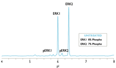
Simple Western Charge Data
Simple Western Charge data is processed automatically in Compass for Simple Western software. Sample data is displayed as an electropherogram where it's easier to see minute changes. Quantitative results such as pI value, signal intensity (area), % area, and signal-to-noise for each immunodetected protein are presented in the results table automatically.
From Your Peers Using Simple Western Charge
“The Simple Western Charge assay, a capillary isoelectric focusing immunoassay, exceeded the reliability of 2D Western blots for resolving recombinant PKG-Iα and PKG-Iβ and uses 100,000X less sample quantity, making it well-suited for clinical disease proteomics because protein isoforms and post-translational modifications can be detected in precious tissue biopsies.”
- Mary G. Johlfs, M.S., Director of Research Operations/Scientist, Roseman University of Health Sciences

Charge Your Research with Simple Western
The ability to detect and quantify nearly all protein isoforms with high sensitivity and resolution makes Simple Western Charge a uniquely powerful method for studying cell signaling networks, cancer, among many
other applications.
This eBook provides protocols for the analysis of more than 30 proteins by Simple Western Charge, including common components of signaling pathways, enzymes, membrane proteins, and more.

Enabling Cutting Edge Biomedical Research
Characterizing the charge heterogeneity of biomolecules can provide profound insight into biological processes. For example, a protein’s charge heterogeneity will change in response to post-translational modifications like phosphorylation, and these changes may not be readily detectable by size-based assays like the traditional Western blot.
Simple Western Charge assays have been used extensively in biomedical research, creating charge heterogeneity fingerprints used to study topics related to cancer, neurological disease, diabetes and more.

Detailed Mapping of Signaling Cascades
Learn how researchers at Manchester University have developed. assays encompassing clinical samples from Chronic Myeloid Leukaemia (CML-SC), Chronic Lymphocytic Leukaemia (CLL), Non-Small Cell Lung Cancer (NSCLC; tissue & plasma) and Endometrial Cancer using the Simple Western Charge assay. This work defines the utility of the Simple Western Charge assay in detailed mapping of signaling cascades in model systems and cellular organelles.
