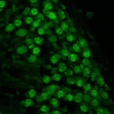
Immunohistochemical Analysis of 53BP1 in Paraffin Embedded Breast Tumors
Human breast tumors stained with 53BP1 antibody. Image from verified customer review.
53BP1 Staining in Multiple Tissues
Detection of Human and Mouse 53BP1 by IHC. Sample: FFPE sections of human ovarian carcinoma (left) and mouse teratoma (right). Antibody: 53BP1 Antibody (Catalog #NB100-304) used at a dilution of 1:1000 (1ug/mL). Detection: DAB.
Immunocytochemistry/Immunofluorescence Staining of 53BP1 in Multiple Cell Lines
Human medulloblastoma (DAOY) and mouse astrocyte (C8D1A) cell lines were exposed for 48 hours to DMSO or 1ug/mL of the DNA damaging agent EPE. Cells were immunostained for 53BP1 (green). The nuclei were counterstained with DAPI (blue). Image from verified customer review.
Intracellular Staining of 53BP1 in RH-30 Cells in Flow Cytometry
An intracellular stain was performed on RH-30 cells with 53BP1 Antibody (Catalog #NB100-304AF647) (blue) and a matched isotype control (orange). Cells were fixed with 4% PFA and then permeabilized with 0.1% saponin. Cells were incubated in an antibody dilution of 5 ug/mL for 30 minutes at room temperature. Both antibodies were conjugated to Alexa Fluor 647.
Detection of 53BP1 in Immunoprecipitated HCC44 Cell Lysate
Immunoprecipitation analysis lysates from HCC44 cells in 1% NP40. Image from verified customer review.
Fluorescent Detection of 53BP1 in Irradiated A549 Cells
Effect of RK-33 on radiation-induced DNA damage. A Immunofluorescence images showing 53BP1 and gamma-H2AX foci in A549 cells after 2-Gy radiation and A549 cells pre-treated with 6 uM RK-33, 12 h before radiation. Overlap of 53BP1 and gamma-H2AX is seen in the merged picture of the co-immunofluorescence staining. Scale bar is 2 um. Image collected and cropped by CiteAb from the following publication (http://embomolmed.embopress.org/cgi/doi/10.15252/emmm.201404368) licensed under a CC-BY licence.
Western Blot of 53BP1 in U2OS Cells Exposed to Ionizing Radiation
Lamin A/C-53BP1 interaction is regulated in a DNA damage-dependent manner. U2OS/GFP-lamin A cells were pretreated with 10 um ATMi f for 1 h before exposure to IR (10 Gy, 1 h recovery). Cell extracts were then subjected to immunoprecipitation using GFP-Trap beads, and bound complexes were then analyzed by immunoblotting using 53BP1 and GFP antibodies. WCE represents 1% input. Image collected and cropped by CiteAb from the following publication (http://doi.wiley.com/10.1111/acel.12258), licensed under a CC-BY licence.
Immunocytochemistry/Immunofluorescence Staining of 53BP1 in HeLa Cells
HeLa cells were fixed in 4% paraformaldehyde for 10 minutes and permeabilized in 0.5% Triton X-100 in PBS for 5 minutes. The cells were incubated with 53BP1 Antibody conjugated to DyLight 550 (NB100-304R) at 5 ug/ml for 1 hour at room temperature. Nuclei were counterstained with DAPI (Blue). Cells were imaged using a 40X objective.
Intracellular Staining of 53BP1 in Ntera2 Cells in Flow Cytometry
An intracellular stain was performed on Ntera2 cells with 53BP1 Antibody NB100-304 (blue) and a matched isotype control NBP2-24891 (orange). Cells were fixed with 4% PFA and then permeabilized with 0.1% saponin. Cells were incubated in an antibody dilution of 1.0 ug/mL for 30 minutes at room temperature, followed by Rabbit IgG (H+L) Cross-Adsorbed Secondary Antibody, Dylight 550 (SA5-10033, Thermo Fisher).
Western Blot of 53BP1 in Multiple Cell Lines
Whole cell lysate from U2OS or 293T cells. Bands indicate an observed molecular weight of ~220 kDa and the theoretical molecular weight is 214 kDa.
Immunocytochemistry/Immunofluorescence of Irradiated and Non-Irradiated MEFs
Upper Panel: 53BP1 foci in proliferating MEFs. Lower Panel: 53BP1 foci in proliferating MEFs exposed to 10 Gy of IR.
Immunocytochemistry/Immunofluorescence Staining of 53BP1 in Embryonic Fibroblast Cells
Embryonic Fibroblast cells pre-extraction for 5 mins with CSK buffer. Fixed with 4% PFA and 75% Ethanol. Primary Antibody at 1:1000. Secondary Antibody at 1:1000. Image from verified customer review.
Probing of 53BP1 in Untreated and Irradiated Cells
Upper Panel: Control untreated cells. Lower Panel: Cells exposed to irradiation (10 Gy) and probed for 53BP1 foci. Cells were grown on coverslips, fixed with 4% paraformaldehyde, methanol permeabilized, blocked for 1 h, RT. Incubated with primary antibody (1:200) overnight, washed 3x with PBS, probed with tubulin (Alexa Fluor 594) antibody for 2 h, RT. Washed 3x with PBS, mounted on slides using prolong gold, imaged using Nikon confocal microscope (100x oil). Image from verified customer review.
Immunocytochemistry/Immunofluorescence Staining of 53BP1 in Transfected H1299 Cells
MUS81 inhibition in BRCA2-deficient cells causes accumulation of 53BP1 nuclear bodies and G1 arrest. H1299 cells carrying a DOX-inducible BRCA2 shRNA were transfected with control or MUS81 siRNAs. Representative images of cells processed 72 h later for immunostaining with 53BP1 Antibody (green) and anti-cyclin A (red) antibodies. DNA was counterstained with DAPI. Scale bar, 10 um. Image collected and cropped by CiteAb from the following publication (http://www.nature.com/doifinder/10.1038/ncomms15983), licensed under a CC-BY licence.
Immunocytochemistry/Immunofluorescence of 53BP1 in Control and shPot1a-1 Transduced LSK Cells
Pot1a prevents DDR in HSCs. Telomeric DDR in 8 week-old LSK cells upon Pot1a knockdown. Immunocytochemical staining of TRF1 (green). Foci co-stained with TRF1 and 53BP1 were identified as TIFs. Nuclei were stained with TOTO3 (blue). Scale bar, 2 um. Image collected and cropped by CiteAb from the following publication (http://www.nature.com/articles/s41467-017-00935-4), licensed under a CC-BY licence.
Immunofluorescence of 53BP1 in Transfected U2OS Cells
Lamin A/C-53BP1 interaction is regulated in a DNA damage-dependent manner. U2OS cells were transfected with siCTRL or siLMNA and subjected to laser micro-irradiation, fixed 15 min later and then processed for immunofluorescence with gamma-H2AX and 53BP1 antibodies. Scale bar, 10 um. Image collected and cropped by CiteAb from the following publication (http://doi.wiley.com/10.1111/acel.12258), licensed under a CC-BY licence.
Immunofluorescent Staining of 53BP1 in Ntera2 Cells
Ntera2 cells were fixed in 4% paraformaldehyde for 10 minutes and permeabilized in 0.5% Triton X-100 in PBS for 5 minutes. The cells were incubated with anti-53BP1 Antibody NB100-304 at 2 ug/ml overnight at 4C and detected with an anti-rabbit Dylight 488 (Green) at a 1:1000 dilution for 60 minutes. Alpha tubulin (DM1A) NB100-690 was used as a co-stain at a 1:1000 dilution and detected with an anti-mouse Dylight 550 (Red) at a 1:1000 dilution. Nuclei were counterstained with DAPI (Blue). Cells were imaged using a 100X objective and digitally deconvolved.
Immunofluorescent Staining of 53BP1 in NIH3T3 Cells
NIH3T3 cells were fixed in 4% paraformaldehyde for 10 minutes and permeabilized in 0.5% Triton X-100 in PBS for 5 minutes. The cells were incubated with anti-53BP1 Antibody NB100-304 at 2 ug/ml overnight at 4C and detected with an anti-rabbit Dylight 488 (Green) at a 1:1000 dilution for 60 minutes. Alpha tubulin (DM1A) NB100-690 was used as a co-stain at a 1:1000 dilution and detected with an anti-mouse Dylight 550 (Red) at a 1:1000 dilution. Nuclei were counterstained with DAPI (Blue). Cells were imaged using a 100X objective and digitally deconvolved.
Immunocytochemistry/Immunofluorescence in HeLa Cells Using Conjugated 53BP1 Antibody
HeLa cells were fixed in 4% paraformaldehyde for 10 minutes and permeabilized in 0.5% Triton X-100 in PBS for 5 minutes. The cells were incubated with 53PB1 Antibody conjugated to DyLight 550 (NB100-304R) at 5 ug/ml for 1 hour at room temperature. Nuclei were counterstained with DAPI (Blue). Cells were imaged using a 100X objective and digitally deconvolved.
Immunohistochemical Staining of 53BP1 in Paraffin Embedded Colon Cancer
Staining of 53BP1 in human colon cancer using DAB with hematoxylin counterstain.
Flow Cytometry of A431 Cells Stained with Conjugated 53BP1 Antibody
An intracellular stain was performed on A431 cells with 53BP1 Antibody (Catalog #NB100-304AF647) (blue) and a matched isotype control (orange). Cells were fixed with 4% PFA and then permeabilized with 0.1% saponin. Cells were incubated in an antibody dilution of 2.5 ug/mL for 30 minutes at room temperature. Both antibodies were conjugated to Alexa Fluor 647.
Cells Stained with 53BP1 Antibody in Immunocytochemistry/Immunofluorescence
For 53BP1 staining, samples were incubated for 1 hour with 1:200 53BP1 primary antibody in PBS/BSA 1%, at room temperature. Green: 53BP Blue: DAPI. Image from verified customer review.


