53BP1 Antibody [Alexa Fluor® 488]

Novus Biologicals, part of Bio-Techne | Catalog # NB100-304AF488

Novus Biologicals, part of Bio-Techne

Key Product Details

Species Reactivity

Validated:

Human, Mouse, Rat, Bat, Bovine, Canine, Fish, Goat, Primate

Predicted:

Chimpanzee (96%), Feline (90%), Orangutan (96%), Porcine (90%), Rabbit (90%), Sheep (90%). Backed by our 100% Guarantee.

Applications

Flow (Intracellular), Flow Cytometry, Immunoblotting, Immunocytochemistry/Immunofluorescence, Immunohistochemistry, Immunohistochemistry-Frozen, Immunohistochemistry-Paraffin, Knockdown Validated, Knockout Validated, Western Blot

Label

Alexa Fluor 488 (Excitation = 488 nm, Emission = 515-545 nm)

Antibody Source

Polyclonal Rabbit IgG

Concentration

Please see the vial label for concentration. If unlisted please contact technical services.

Product Specifications

Immunogen

Partial synthetic peptide made to an internal portion of human 53BP1 (between amino acids 350-400) [NP_005648.1].

Reactivity Notes

Human reactivity reported in scientific literature (PMID:32877678). Human and mouse reactivity cited in numerous publications. Primate reactivity reported in scientific literature (PMID: 18389475). Fish reactivity reported in scientific literature (PMID: 25516420). Bat, canine, and bovine reactivity reported in scientific literature (PMID: 27573809). Predicted cross-reactivity based on sequence identity: Chimpanzee (96%), Gorilla (96%), Orangutan (96%), Gibbon (94%), Marmoset (92%), Feline (90%), Porcine (90%), Rabbit (90%), Sheep (90%).

Marker

DNA Double Strand Break Marker

Clonality

Polyclonal

Host

Rabbit

Isotype

IgG

Scientific Data Images for 53BP1 Antibody [Alexa Fluor® 488]

Immunocytochemistry/Immunofluorescence Using Conjugated 53BP1 Antibody

Immunocytochemistry/Immunofluorescence Using Conjugated 53BP1 Antibody

53BP1 Antibody [Alexa Fluor 488] [NB100-304AF488]
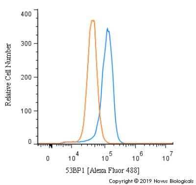
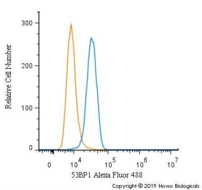
Flow Cytometry of NIH3T3 Cells Stained with Conjugated 53BP1 Antibody

Flow Cytometry of NIH3T3 Cells Stained with Conjugated 53BP1 Antibody

An intracellular stain was performed on NIH3T3 cells with 53BP1 Antibody (Catalog #NB100-304AF488) (blue) and a matched isotype control (orange). Cells were fixed with 4% PFA and then permeabilized with 0.1% saponin. Cells were incubated in an antibody dilution of 5 ug/mL for 30 minutes at room temperature. Both antibodies were conjugated to Alexa Fluor 488.
Immunocytochemistry/Immunofluorescence in HeLa Cells Using Conjugated 53BP1 Antibody

Immunocytochemistry/Immunofluorescence in HeLa Cells Using Conjugated 53BP1 Antibody

HeLa cells were fixed for 10 minutes using 10% formalin and then permeabilized for 5 minutes using 1X PBS + 0.5% Triton-X100. The cells were incubated with 53BP1 Antibody conjugated to Alexa Fluor 488 (Catalog #NB100-304AF488) at 10 ug/ml for 1 hour at room temperature. Nuclei were counterstained with DAPI (Blue). Cells were imaged using a 40X objective.

Applications for 53BP1 Antibody [Alexa Fluor® 488]

Application
Recommended Usage

Flow (Intracellular)

Optimal dilutions of this antibody should be experimentally determined.

Flow Cytometry

Optimal dilutions of this antibody should be experimentally determined.

Immunoblotting

Optimal dilutions of this antibody should be experimentally determined.

Immunocytochemistry/Immunofluorescence

Optimal dilutions of this antibody should be experimentally determined.

Immunohistochemistry

Optimal dilutions of this antibody should be experimentally determined.

Immunohistochemistry-Frozen

Optimal dilutions of this antibody should be experimentally determined.

Immunohistochemistry-Paraffin

Optimal dilutions of this antibody should be experimentally determined.

Knockdown Validated

Optimal dilutions of this antibody should be experimentally determined.

Knockout Validated

Optimal dilutions of this antibody should be experimentally determined.

Western Blot

Optimal dilutions of this antibody should be experimentally determined.
Application Notes
Optimal dilution of this antibody should be experimentally determined.

Reviewed Applications

Read 1 review rated 5 using NB100-304AF488 in the following applications:

Formulation, Preparation, and Storage

Purification

Immunogen affinity purified

Formulation

50mM Sodium Borate

Preservative

0.05% Sodium Azide

Concentration

Please see the vial label for concentration. If unlisted please contact technical services.

Shipping

The product is shipped with polar packs. Upon receipt, store it immediately at the temperature recommended below.

Stability & Storage

Store at 4C in the dark.

Background: 53BP1

Tumor protein p53 binding protein 1 (P53-binding protein 1 or 53BP1) plays a critical role in tumor suppression and is a putative substrate of ATM kinase with a theoretical molecular weight of 214 kDa. Upon DNA damage, it is phosphorylated and relocalizes to the presumptive sites of damage, specifically, double-strand breaks. 53BP1 plays a key role in response to DNA damage, acts as a signaling checkpoint during mitosis, and enhances TP53-mediated transcriptional activation. Originally identified as p53's transcriptional enhancing partner, 53BP1 is known as a key substrate for ataxia telangiectasia mutated (ATM) signaling, whose function to generate gamma H2AX may be partially compensated by the activity of DNA-dependent kinase (DNA-PK). 53BP1 relocalizes to discrete foci overlapping with gamma phosphorylated histone H2AX; demarcating DNA double strand breaks (DSBs) sites following exposure to radiation (1). 53BP1 functions downstream of gamma H2AX-dependent proteins that collectively establish ionizing radiation induced foci at DSBs. 53BP1 is downstream of Mre11/Rad50/NBS1 (MRN complex), MDC1, RNF8, RNF168 and HERC2 which recruit 53BP1 to the DSB site, suggesting a role in DNA repair through genomic stability maintenance (2).

References

1.Henry, E., Souissi-Sahraoui, I., Deynoux, M., Lefevre, A., Barroca, V., Campalans, A., . . . Arcangeli, M. L. (2019). Human hematopoietic stem/progenitor cells display ROS-dependent long-term hematopoietic defects after exposure to low dose of ionizing radiations. Haematologica. doi:10.3324/haematol.2019.226936

2.Janoshazi, A. K., Horton, J. K., Zhao, M. L., Prasad, R., Scappini, E. L., Tucker, C. J., & Wilson, S. H. (2020). Shining light on the response to repair intermediates in DNA of living cells. DNA Repair (Amst), 85, 102749. doi:10.1016/j.dnarep.2019.102749

Long Name

p53 Binding Protein 1

Alternate Names

p202, TP53BP1

Gene Symbol

TP53BP1

Additional 53BP1 Products

Product Documents for 53BP1 Antibody [Alexa Fluor® 488]

Certificate of Analysis

To download a Certificate of Analysis, please enter a lot number in the search box below.

Product Specific Notices for 53BP1 Antibody [Alexa Fluor® 488]

Alexa Fluor (R) products are provided under an intellectual property license from Life Technologies Corporation. The purchase of this product conveys to the buyer the non-transferable right to use the purchased product and components of the product only in research conducted by the buyer (whether the buyer is an academic or for-profit entity). The sale of this product is expressly conditioned on the buyer not using the product or its components, or any materials made using the product or its components, in any activity to generate revenue, which may include, but is not limited to use of the product or its components: (i) in manufacturing; (ii) to provide a service, information, or data in return for payment; (iii) for therapeutic, diagnostic or prophylactic purposes; or (iv) for resale, regardless of whether they are resold for use in research. For information on purchasing a license to this product for purposes other than as described above, contact Life Technologies Corporation, 5791 Van Allen Way, Carlsbad, CA 92008 USA or outlicensing@lifetech.com. This conjugate is made on demand. Actual recovery may vary from the stated volume of this product. The volume will be greater than or equal to the unit size stated on the datasheet.

This product is for research use only and is not approved for use in humans or in clinical diagnosis. Primary Antibodies are guaranteed for 1 year from date of receipt.
Loading...
Loading...
Loading...
Loading...
Loading...
Loading...