Calnexin Antibody - BSA Free
Novus Biologicals, part of Bio-Techne | Catalog # NB100-1974


Conjugate
Catalog #
Key Product Details
Species Reactivity
Validated:
Human, Mouse, Rat, Hamster, Zebrafish
Cited:
Human, Mouse, Hamster, Zebrafish
Predicted:
Canine (90%), Orangutan (93%). Backed by our 100% Guarantee.
Applications
Validated:
Flow Cytometry, Immunocytochemistry/ Immunofluorescence, Immunohistochemistry, Immunohistochemistry-Paraffin, Immunoprecipitation, Simple Western, Western Blot
Cited:
FLOW, ICC/IF, WB
Label
Unconjugated
Antibody Source
Polyclonal Rabbit IgG
Format
BSA Free
Concentration
1 mg/ml
Product Specifications
Immunogen
A synthetic peptide made to a C-terminal portion of the rat Calnexin protein (between residues 550-591)
Reactivity Notes
Hamster reactivity reported in the scientific literature (PMID: 23760268). Use in Zebrafish reported in scientific literature (PMID:23049555).
Localization
Endoplasmic reticulum
Marker
Endoplasmic Reticulum Membrane Marker
Clonality
Polyclonal
Host
Rabbit
Isotype
IgG
Theoretical MW
97 kDa.
Disclaimer note: The observed molecular weight of the protein may vary from the listed predicted molecular weight due to post translational modifications, post translation cleavages, relative charges, and other experimental factors.
Disclaimer note: The observed molecular weight of the protein may vary from the listed predicted molecular weight due to post translational modifications, post translation cleavages, relative charges, and other experimental factors.
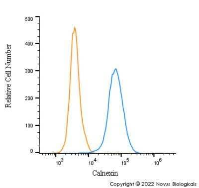
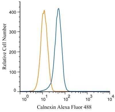
Scientific Data Images for Calnexin Antibody - BSA Free
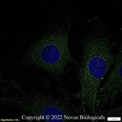
Immunocytochemistry/Immunofluorescence: Calnexin Antibody [NB100-1974] - HeLa cells were fixed in 4% paraformaldehyde for 10 minutes and permeabilized in 0.05% Triton X-100 in PBS for 5 minutes. The cells were incubated with (NB100-1974) at 1 ug/ml overnight at 4C and detected with an anti-rabbit DyLight 488 (Green) at a 1:1000 dilution for 60 minutes. Nuclei were counterstained with DAPI (Blue). Cells were imaged using a 100X objective and digitally deconvolved.
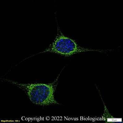
Immunocytochemistry/Immunofluorescence: Calnexin Antibody [NB100-1974] - NIH3T3 cells were fixed in 4% paraformaldehyde for 10 minutes and permeabilized in 0.05% Triton X-100 in PBS for 5 minutes. The cells were incubated with (NB100-1974) at 1 ug/ml overnight at 4C and detected with an anti-rabbit DyLight 488 (Green) at a 1:1000 dilution for 60 minutes. Nuclei were counterstained with DAPI (Blue). Cells were imaged using a 100X objective and digitally deconvolved.
Immunocytochemistry/Immunofluorescence: Calnexin Antibody [NB100-1974] - Rat FR cells were fixed in 4% paraformaldehyde for 10 minutes and permeabilized in 0.05% Triton X-100 in PBS for 5 minutes. The cells were incubated with (NB100-1974) at 1 ug/ml overnight at 4C and detected with an anti-rabbit DyLight 488 (Green) at a 1:1000 dilution for 60 minutes. Nuclei were counterstained with DAPI (Blue). Cells were imaged using a 100X objective and digitally deconvolved.
Applications for Calnexin Antibody - BSA Free
Application
Recommended Usage
Immunocytochemistry/ Immunofluorescence
1:100
Immunohistochemistry
1:100
Immunohistochemistry-Paraffin
1:100
Immunoprecipitation
1:100
Simple Western
1:50
Western Blot
1:1000
Application Notes
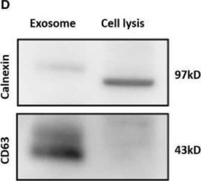
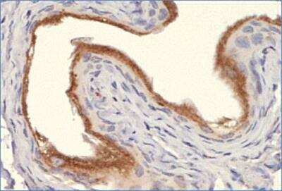
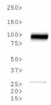
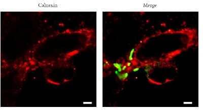
In ICC/IF, endoplasmic reticulum staining was observed in HeLa cells. In Western Blot, a band is seen at ~ 90 kDa representing Calnexin. In IHC-P, staining was observed in the endoplasmic reticulum of mouse bladder tissue. Prior to immunostaining paraffin tissues, antigen retrieval with sodium citrate buffer (pH 6.0) is recommended. In Simple Western only 10 - 15 uL of the recommended dilution is used per data point. Separated by Size-Wes, Sally Sue/Peggy Sue.
Reviewed Applications
Read 1 review rated 4 using NB100-1974 in the following applications:
Formulation, Preparation, and Storage
Purification
Immunogen affinity purified
Formulation
PBS
Format
BSA Free
Preservative
0.02% Sodium Azide
Concentration
1 mg/ml
Shipping
The product is shipped with polar packs. Upon receipt, store it immediately at the temperature recommended below.
Stability & Storage
Store at 4C short term. Aliquot and store at -20C long term. Avoid freeze-thaw cycles.
Background: Calnexin
Alternate Names
calnexin, CNX, IP90FLJ26570, Major histocompatibility complex class I antigen-binding protein p88, p90
Gene Symbol
CANX
UniProt
Additional Calnexin Products
Product Documents for Calnexin Antibody - BSA Free
Product Specific Notices for Calnexin Antibody - BSA Free
This product is for research use only and is not approved for use in humans or in clinical diagnosis. Primary Antibodies are guaranteed for 1 year from date of receipt.
Loading...
Loading...
Loading...
Loading...
Loading...


![Western Blot: Calnexin Antibody - BSA Free [NB100-1974] - Calnexin Antibody - BSA Free](https://resources.bio-techne.com/images/products/nb100-1974_rabbit-polyclonal-calnexin-antibody-291220231213292.jpg)
![Western Blot: Calnexin Antibody - BSA Free [NB100-1974] - Calnexin Antibody - BSA Free](https://resources.bio-techne.com/images/products/nb100-1974_rabbit-polyclonal-calnexin-antibody-2322024894227.jpg)
![Western Blot: Calnexin Antibody - BSA Free [NB100-1974] - Calnexin Antibody - BSA Free](https://resources.bio-techne.com/images/products/nb100-1974_rabbit-polyclonal-calnexin-antibody-23220248135260.jpg)
![Western Blot: Calnexin Antibody - BSA Free [NB100-1974] - Calnexin Antibody - BSA Free](https://resources.bio-techne.com/images/products/nb100-1974_rabbit-polyclonal-calnexin-antibody-2322024822139.jpg)
![Western Blot: Calnexin Antibody - BSA Free [NB100-1974] - Calnexin Antibody - BSA Free](https://resources.bio-techne.com/images/products/nb100-1974_rabbit-polyclonal-calnexin-antibody-23220248194398.jpg)
