Caveolin-1 Antibody (7C8)
Novus Biologicals, part of Bio-Techne | Catalog # NB100-615


Conjugate
Catalog #
Forumulation
Catalog #
Key Product Details
Species Reactivity
Validated:
Human, Mouse, Rat, Sheep
Cited:
Human, Mouse, Rat, Insect - Drosophila melanogaster, Ovine
Applications
Validated:
Flow Cytometry, Immunoblotting, Immunocytochemistry/Immunofluorescence, Immunohistochemistry, Immunohistochemistry-Frozen, Immunohistochemistry-Paraffin, Immunoprecipitation, Western Blot
Cited:
Flow Cytometry, IF/IHC, Immunoblotting, Immunocytochemistry/ Immunofluorescence, Immunohistochemistry, Immunohistochemistry-Frozen, Immunohistochemistry-Paraffin, Immunoprecipitation, Western Blot
Label
Unconjugated
Antibody Source
Monoclonal Mouse IgG2B Clone # 7C8
Concentration
1.0 mg/ml
Product Summary for Caveolin-1 Antibody (7C8)
Immunogen
Intracellular membrane protein-containing vesicles (containing GLUT4) from rat adipocytes
Localization
Cell Membrane
Specificity
This is specific for caveolin alpha and beta proteins.
Marker
Caveolae Marker, Endosome Marker
Clonality
Monoclonal
Host
Mouse
Isotype
IgG2B
Theoretical MW
23 kDa.
Disclaimer note: The observed molecular weight of the protein may vary from the listed predicted molecular weight due to post translational modifications, post translation cleavages, relative charges, and other experimental factors.
Disclaimer note: The observed molecular weight of the protein may vary from the listed predicted molecular weight due to post translational modifications, post translation cleavages, relative charges, and other experimental factors.
Scientific Data Images for Caveolin-1 Antibody (7C8)
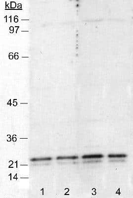
Western Blot: Caveolin-1 Antibody (7C8) [NB100-615] - Detection of caveolin in 3T3 cell lysates (50 ug). Lanes 1 and 2: 1:4000. Lanes 3 and 4: 1:1000. Detection by ECL: 5 minute exposure.

Immunocytochemistry/Immunofluorescence: Caveolin-1 Antibody (7C8) [NB100-615] - Subcellular localization of mPARM-1 and hPARM-1 (full-length and mutant proteins). NIH/3T3 cells transiently expressing hPARM-1-GFP or deltaCT-GFP were fixed, immunostained for caveolin-1 (1:100, Novus Biologicals), and examined by confocal fluorescence microscopy. For hPARM-1-GFP-caveolin-1 co-localization, cells that demonstrated cell membrane PARM-1 localization were chosen. All co-localizations were observed following merging images of GFP-tagged proteins with those of Golgi, endosomes, plasma membrane, alpha-tubulin or caveolin-1 labeling. Image collected and cropped by CiteAb from the following publication (http://molecular-cancer.biomedcentral.com/articles/10.1186/1476-4598-12-84), licensed under a CC-BY license.

Immunohistochemistry-Frozen: Caveolin-1 Antibody (7C8) [NB100-615] - Immunofluorescence of human adipose tissue. Primary antibody at 1:250. IHC-Fr image submitted by a verified customer review.


Applications for Caveolin-1 Antibody (7C8)
Application
Recommended Usage
Flow Cytometry
1 ug per million cells
Immunoblotting
reported in scientific literature (PMID 31759628)
Immunocytochemistry/Immunofluorescence
1:200
Immunohistochemistry
1:100-1:300
Immunohistochemistry-Frozen
reported in scientific literature (PMID 24758774)
Immunohistochemistry-Paraffin
1:100-1:300
Immunoprecipitation
1-2 ug / 500 ug of protein
Western Blot
1:1000-1:4000
Application Notes
In Western blot, a band is observed ~23 kDa, representing the Caveolin 1 protein. A band at ~21 kDa may also be observed depending on the lysates used.
Please Note: Optimal dilutions of this antibody should be experimentally determined.
Reviewed Applications
Read 3 reviews rated 5 using NB100-615 in the following applications:
Formulation, Preparation, and Storage
Purification
Protein A purified
Formulation
Tris-Glycine, 0.15 M NaCl
Preservative
0.05% Sodium Azide
Concentration
1.0 mg/ml
Shipping
The product is shipped with polar packs. Upon receipt, store it immediately at the temperature recommended below.
Stability & Storage
Aliquot and store at -20C or -80C. Avoid freeze-thaw cycles.
Background: Caveolin-1
Alternate Names
CAV1, Caveolin1, MSTP085, VIP21
Gene Symbol
CAV1
Additional Caveolin-1 Products
Product Documents for Caveolin-1 Antibody (7C8)
Product Specific Notices for Caveolin-1 Antibody (7C8)
This product is for research use only and is not approved for use in humans or in clinical diagnosis. Primary Antibodies are guaranteed for 1 year from date of receipt.
Loading...
Loading...
Loading...
Loading...
Loading...
Loading...